Worms Found During Bladder Expression

Blacky, a 3-year-old, female mixed-breed dog, presented for preoperative evaluation prior to scheduled ovariohysterectomy.
Physical Examination
The dog’s body temperature, heart rate, and respiratory rate were within normal limits. No abnormal physical examination findings were noted.
Preoperative Diagnostics
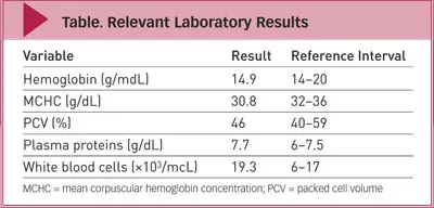
Routine preoperative laboratory analysis included a complete blood count (see Table), which revealed mild deviations in red blood cell, white blood cell, and plasma protein parameters.
An enzyme-linked immunosorbent assay (ELISA) antigen test (SNAP 3Dx, idexx.com) to survey for canine infectious diseases was positive for heartworm. However, no microfilariae were observed in a wet mount. During bladder expression, 2 worms were eliminated in the dog’s urine. No parasite eggs were noted in urine sediment.

Ask Yourself…
Which common nematodes can be found in the kidney and bladder of a dog?
Describe the clinical signs associated with parasites of the urinary system.
DIAGNOSIS: Heartworm Disease
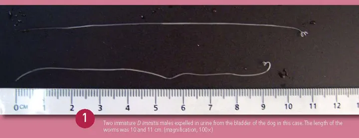
The 2 worms found in the urine of this dog were identified as immature male Dirofilaria immitis nematodes, measuring 10 and 11 cm (Figure 1). Type, sex, and stage were determined morphologically; males have coiled tails while females have tapered, straight tails. The length confirmed that they were immature worms.
PathogenesisD immitis is a pathogenic filarial nematode that infects many mammals, including dogs and cats.1 Cutaneous penetration of an infective third-stage larva can result from the bite of a mosquito in a definitive host following a period of development within the mosquito.
In dogs, the larva develops and matures over the next 6 months while migrating to the pulmonary arteries, where it develops into a mature worm. The infection and associated pathology in the pulmonary vasculature, and occasionally right ventricle of the heart, of dogs are referred to as heartworm disease.1
Canine heartworm disease is endemic on the island of Grenada,2 as it is in many tropical regions with large populations of stray dogs and high numbers of susceptible pets that are not maintained on regular heartworm prophylaxis.
Two immature D immitis males expelled in urine from the bladder of the dog in this case. The length of the worms was 10 and 11 cm. (magnification, 100x)

Aberrant MigrationTo the best of our knowledge, this is the first report of D immitis worms expelled in urine expressed from the bladder of a dog in Grenada, West Indies (see D immitis Aberrant Migration).
Dogs with aberrantly located adult heartworms have been reported since 1856,3 but the mechanism for aberrant location of heartworms has not been determined.4 However, 1 report documented microfilariae in the urine sediment of a heartworm-positive dog in Thailand,5 and a separate report suggested that glomerular capillaries played a role as a reservoir of Onchocerca volvulus,6 a parasite with microfilariae closely related to D immitis microfilariae.
Histologic examination of the kidneys of dogs with heartworm infection accompanied by mild-to-moderate proteinuria can reveal the presence of microfilariae in the glomerular capillaries and the medullary vessels. Mild-to-severe, diffuse, chronic interstitial nephritis and generalized membranoproliferative glomerulonephritis can also be found.7
Case InterpretationThis dog had not been receiving routine heartworm prophylaxis. The hematology results indicated mild hyperproteinemia and leukocytosis. The observed leukocytosis may have been due to eosinophilia, a common observation in dogs with heartworm disease. Unfortunately, an eosinophil count was not provided in the presurgical hematology report.
D immitis Aberrant Migration
Aberrant migration of D immitis is not very common. However, the authors have reported immature female D immitis nematodes (Figure 2) that were recovered from the peritoneal cavity of 2 dogs.9,10 Since the initial report, there has been 1 additional case of aberrant migration to the peritoneal cavity of a dog.

One immature female D immitis recovered from the peritoneal cavity of a dog during routine spay. Length is 12 to 13 cm. (magnification, 100x)
All the reported cases involved young female adult dogs. None of the dogs was receiving routine heartworm prophylaxis or had clinical signs of heartworm disease. Two of the 3 dogs were positive for heartworms based on ELISA antigen testing. One dog was not tested due to financial constraints of the owner. Diagnosis was based on morphologic identification of the immature worms. All recovered D immitis nematodes were immature males and females.
It has been hypothesized that fourth-stage larvae accessing the arterial blood supply may become lodged in the glomerulus of the kidneys. In the dog presented in this case, it is hypothesized that immature larvae traveled via the arterial circulation to the kidney, where they penetrated into the urinary system via the glomerular capillaries.
No abnormal clinical signs related to heartworm disease were observed. Microfilariae were not observed in the dog’s blood or urine sediment. When D immitis is suspected, however, thoracic radiography is advisable.
Did you answer ...
Capillaria (Pearsonema) plica are thread-like worms found occasionally in the pelvis of the kidney, ureter, or bladder of dogs, cats, and other animals. C plica has been reported in North America, Europe, and Russia.8 Clinical signs of infection are typically absent. Earthworms are the intermediate host; ova appear somewhat similar to those of whipworms (but are colorless with asymmetric bipolar plugs) and are passed in the urine.
The nematode Dioctophyma renale is most commonly seen in the urine of mink but is occasionally found in dogs and other species, including humans.8 Adult D renale are red and live in renal tissues, almost invariably in the right kidney. The renal parenchyma is gradually destroyed as the female worm grows. Pitted, thick-shelled eggs with bipolar plugs are passed in the urine. The intermediate host is an oligochaete (annelid). Infective third-stage larvae may also reside in a paratenic host (eg, frog, fish), which is subsequently ingested by the definitive host.
Clinical presentation is normally inconclusive when diagnosing parasitic infection of the kidneys, ureters, and bladder of dogs. Individuals affected by renal parasites typically have no clinical signs, especially those infested with Capillaria species.10 However, some pets may be extremely ill if they have associated kidney failure or a severe renal infection.10,11 Historical signs of renal infection include hematuria, abdominal pain or painful urination, recurrent urinary tract infections, and frequent urination.10
Clinical signs of heartworm infection do not develop until the parasites approach or reach maturity. Therefore, signs are often historical, not clinical, and might include exercise intolerance or mild weight loss. In addition, heartworm disease should always be considered in endemic areas.
Treatment
The owner declined heartworm treatment, both adulticidal and microfilaricidal, because of financial constraints. However, the ovariohysterectomy was performed without any complications.
The dog was released to the owner but lost to follow-up.
The authors would like to thank the following faculty and graduates from St. George’s University School of Veterinary Medicine, all of whom were involved in the reported cases of aberrant heartworm migration: Dr. L. Telesford, Dr. S. Sage, Dr. C. Fernandez, Dr. Ana-Sophia Pearse, Dr. Michelle Londono, Dr. W. Sylvester, Dr. M. Bhaiyat, Dr. Terra Martin, and Ms. Camille-Marie Coomansingh, MSc.