Vomiting & Diarrhea in a Lethargic Dog
April Summers, DVM, PhD, Cornell University
Julien Guillaumin, DVM, DACVECC, DECVECC, The Ohio State University
Max, an 8-year-old, 32-lb (14.5-kg), neutered male border collie, was emergently presented for vomiting, diarrhea, and lethargy of 3 days’ duration. He was fed a veterinary commercial diet but, when he began vomiting, was transitioned to a bland diet of boneless, skinless boiled chicken and white rice; however, he continued to vomit while on the bland diet.
Max had a previous history of 2 exploratory laparotomies for foreign bodies but had no other major medical history. He was the only dog in the household, had access to a fenced backyard, and had no significant travel history. He was up to date on vaccines and flea, tick, and heartworm prevention.
Physical Examination
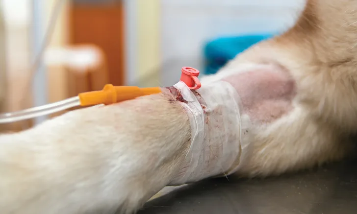
On presentation, Max was laterally recumbent, obtunded, and ≈7% dehydrated. He was pyrexic (temperature, 104.5°F [40.3°C]) and tachycardic (150 bpm) with a normal respiratory rate (28 breaths per minute). Mucous membranes were pale, and capillary refill time was prolonged (ie, 3 seconds). He had weak peripheral pulses, and his distal limbs and paw pads were cool to the touch. Harsh lung sounds were auscultated bilaterally throughout all lung fields. He was painful on abdominal palpation, particularly in the cranial abdomen, and had profuse hemorrhagic diarrhea; he also passed pieces of 2 rubber ball toys from his rectum. His owners did not report any foreign material having been passed at home. The remainder of the physical examination was unremarkable.
VENOUS BLOOD GAS RESULTS
Diagnosis & Clinical Management
Venous blood gas analysis revealed metabolic acidosis due to hyperlactatemia and hypoglycemia (Table 1). ECG revealed sinus tachycardia. Systolic blood pressure was 40 mm Hg (Doppler); an arterial catheter was subsequently placed, and direct arterial blood pressure monitoring was initiated.
Images obtained using abdominal-focused assessment with sonography for trauma revealed no evidence of free fluid. Images obtained using thoracic-focused assessment with sonography for trauma revealed 4 to 5 B lines in the region of the right middle lung lobe; B lines are reverberation artifacts and can be present with interstitial edema as air content decreases. Abdominal radiographs revealed fluid-filled loops of intestine consistent with diffuse ileus; however, there was no evidence of GI obstruction. Abdominal sonograms were suggestive of pancreatitis with focal peritonitis but were otherwise unremarkable. Thoracic radiographs revealed an alveolar pattern in the right middle lung lobe that was suggestive of aspiration pneumonia.
Initial laboratory values revealed leukocytosis characterized by monocytosis and neutrophilia with a left shift (Table 2). Max was also thrombocytopenic. Serum chemistry profile revealed decreased albumin and globulin and increased ALT, ALP, and total bilirubin (Table 3).
CBC RESULTS
Max was stabilized with crystalloid fluid boluses (lactated Ringer’s solution [total, 40 mL/kg IV]), followed by a colloid bolus (6% hydroxyethyl starch in sodium chloride [5 mL/kg IV]) administered over 20 minutes. Of note, the use of synthetic colloids is controversial in humans with sepsis due to increased risk for acute kidney injury. Two 25% dextrose boluses (1 mL/kg IV) were also administered to help treat hypoglycemia. Broad-spectrum antibiotic therapy (ie, ampicillin/sulbactam [30 mg/kg IV], enrofloxacin [10 mg/kg IV]) was initiated within an hour of admission, and oxygen supplementation (100 mL/kg/min increased to a maximum of 4-5 L/min) was provided via nasal cannula.
Because Max’s blood pressure did not respond adequately to fluid therapy and had an oscillometric mean of 50 mm Hg, septic shock was diagnosed. Norepinephrine at 0.2 μg/kg/min CRI was initiated and increased incrementally by 0.1 μg/kg/min to 0.8 μg/kg/min within 45 minutes, at which point vasopressin (0.5 milliUnits/kg/min CRI IV) was initiated.
SERUM CHEMISTRY RESULTS
DIAGNOSIS:
SEPTIC SHOCK WITH SUSPECTED CRITICAL ILLNESS-RELATED CORTICOSTEROID INSUFFICIENCY (CIRCI)
Due to the lack of improvement in blood pressure with increasing doses of vasopressors, CIRCI was strongly suspected. Resting cortisol was 6.96 μg/dL (reference range, 1.8-9 μg/dL), and hydrocortisone sodium succinate (1 mg/kg IV bolus followed by CRI at 0.08 mg/kg/hr) was subsequently initiated to treat possible comorbid CIRCI (see Treatment at a Glance).
TREATMENT AT A GLANCE
Vital parameters and blood work abnormalities should be closely monitored.
Hypoglycemia and electrolyte abnormalities should be treated as necessary.
Blood pressure monitoring and response to vasopressor therapy is an important component of identifying potential CIRCI patients.
Due to the lack of dosage information available for injectable hydrocortisone,7 hydrocortisone at 1 mg/kg IV bolus followed by CRI at 0.08 mg/kg/hr can be considered as a modification of the human protocol.5,8
If injectable hydrocortisone is unavailable, other corticosteroids, including prednisone (0.7-1.4 mg/kg/day), can be considered.
Prognosis & Outcome
Despite treatment and supportive care, Max’s status continued to decline and he suffered cardiorespiratory arrest 6 hours after presentation. Autopsy confirmed aspiration pneumonia, gastroenteritis, and pancreatitis.
Septic shock is uncommonly reported in veterinary medicine, and prognosis is poor even with treatment. Reported survival rate in dogs with hypotension severe enough to require vasopressor therapy has been reported to be ≈10% to 20% as compared with a 40% survival rate in humans.1-3 The prognosis for septic shock for humans and dogs with CIRCI is unknown.
Discussion
CIRCI is defined as inadequate corticosteroid activity in relation to the severity of a patient’s illness (see Take-Home Messages).4 CIRCI has been reported to occur in ≈60% of humans with severe sepsis and septic shock.5 The cause of hypothalamic–pituitary–adrenal axis dysfunction is poorly understood. Potential factors of this dysfunction include decreased production of adrenocorticotropic hormone (ACTH), corticotropin-releasing hormone, and cortisol; dysfunction of their associated receptors; adrenal damage from infarction or hemorrhage; adrenal suppression from chronic exogenous glucocorticoid administration; and inflammatory cytokines causing systemic inflammation-associated glucocorticoid resistance. No specific guidelines exist in veterinary medicine for diagnosing or treating CIRCI, and only rare case reports exist. The recommendation for treatment of CIRCI in humans is administration of injectable hydrocortisone, preferably as a constant rate infusion.6
TAKE-HOME MESSAGES
Septic shock is defined as an infection associated with systemic hypotension that occurs despite adequate fluid resuscitation and requires use of vasopressors to maintain a mean arterial blood pressure ≥65 mm Hg.
Treatment of septic shock is complex and often requires continuous assessment and adjustment based on the dynamic needs of the patient. Early source control and administration of appropriate antibiotics is a critical treatment goal for patients with sepsis.
CIRCI is a form of adrenal insufficiency in critically ill patients and should be suspected in patients with vasopressor-refractory hypotension. In humans, it is recommended to initiate hydrocortisone therapy without additional tests when there is vasopressor-refractory hypotension.4
Diagnosis of CIRCI in veterinary medicine can be challenging, as CIRCI has no specific diagnostic criteria; however, it should be suspected in any patient with vasopressor-refractory hypotension.
There has been no agreement on a single test for definitive diagnosis of CIRCI in humans, although a blunted ACTH stimulation test with a Δ-cortisol result <9 µg/dL and/or a resting cortisol level <10 µg/dL have been suggested to be indicative of CIRCI.4,7,8 In veterinary medicine, a study in septic dogs found that a Δ-cortisol level <3 µg/dL obtained from a standard 1-hour ACTH stimulation test using 250 µg of cosyntropin was associated with hypotension and decreased survival.9
Prompt recognition of potential CIRCI is important, as earlier shock resolution can lead to lower mortality in patients with septic shock.10
Early injectable hydrocortisone administration may be considered as a treatment regimen in septic shock patients with vasopressor-refractory hypotension.6,10
ACTH = adrenocorticotropic hormone, CIRCI = critical illness-related corticosteroid insufficiency