Vitamin A Deficiency in Insectivorous Lizards
Thomas H. Boyer, DVM, DABVP (Reptile & Amphibian), Pet Hospital of Penasquitos, San Diego, California
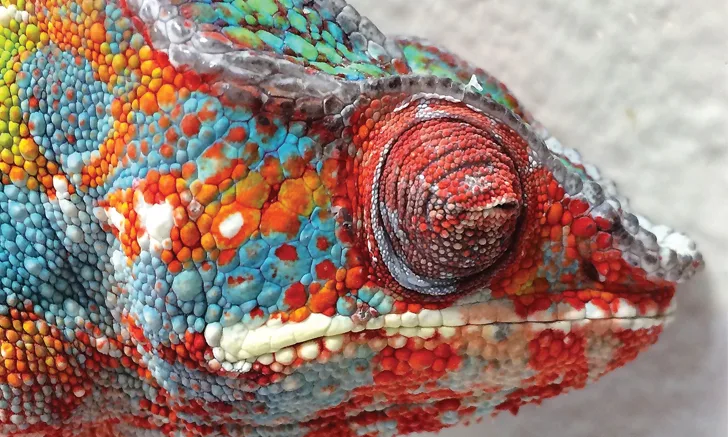
Vitamin A is an essential hormone that activates genes for maturation of immature epidermal cells (ie, keratinocytes). Without vitamin A, cells undergo hyperkeratosis (ie, an abnormal thickening of the stratum corneum, associated with excess keratin) and squamous metaplasia (ie, transformation of cuboidal, columnar, or ciliated glandular or mucosal epithelium into keratinizing stratified squamous epithelium), particularly in the respiratory, ocular, endocrine, GI, and genitourinary systems.1
Background & Pathophysiology
Vitamin A deficiency is common in all captive insectivorous reptiles, including leopard geckos (Eublepharis macularius), chameleons (most commonly panther [Furcifer pardalis], veiled [Chamaeleo calyptratus], and Jackson’s [Trioceros jacksonii]), and anoles. It occurs less often in wild reptiles, likely because of their varied diet, which typically includes other reptiles, mammals, birds, and invertebrates.
Vitamin A deficiency is thought to result from dietary deficiency; it is unknown if insectivorous lizards can synthesize vitamin A from carotenoids in their diet. Dietary history and multivitamin review may disclose a lack of vitamin A in the patient’s diet2; for example, the patient may not be fed multivitamins that contain vitamin A or may be fed insects that do not receive gut-loading diets or other foods containing vitamin A. Feeder insects are generally deficient in vitamin A and often receive diets deficient in vitamin A. In addition, many food manufacturers omit vitamin A and substitute β-carotene in reptile multivitamins because of misinformation that vitamin A is toxic to insectivores.1
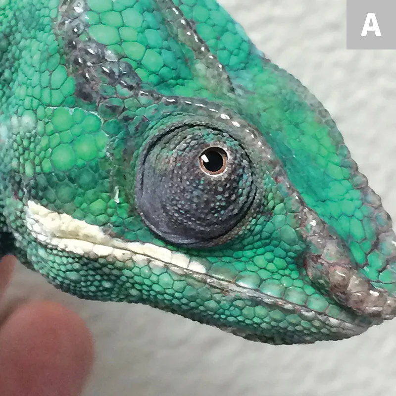
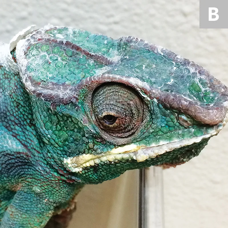
Healthy panther chameleon (Furcifer pardalis; A) with clear rounded eyelids as compared with a panther chameleon with vitamin A deficiency (B). Dull coloration, cheilitis, blepharedema, squinting, mucoid ocular buildup, and dysecdysis are visible over the head and eyelid openings.
Many patients with vitamin A deficiency have long-term ocular disease that is not responsive to multiple antibiotics.2 Both lacrimal and salivary glands are commonly affected, and patients may have blepharitis, blepharospasm (Figure 1), and cheilitis. These patients typically are anorexic and have difficulty shedding (ie, dysecdysis). Leopard geckos may be presented with mucoid-to-solid cellular debris under the eyelids (Figures 2 and 3), ulcerative keratitis, and/or abscessation of periocular glands. Chameleons may have mucoid buildup in the eye, thickened conjunctiva, difficulty capturing prey with the tongue (possibly from dysfunction of sticky mucus glands in the tongue tip), and dull coloration (Figure 4, page 18), which can be difficult to identify on examination. Chronic cases may result in blindness.
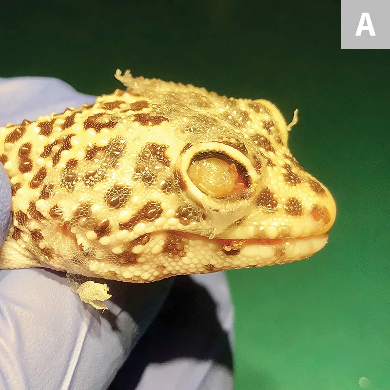
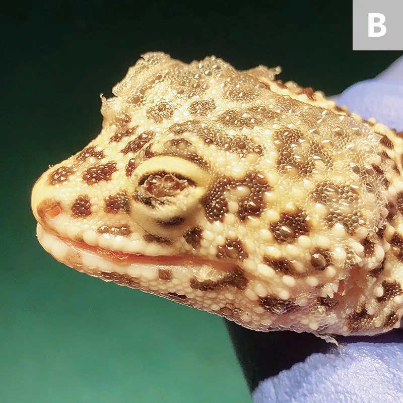
Vitamin A deficiency in a leopard gecko (Eublepharis macularius). Solid cellular debris in the right (A) and left (B) eyes, cheilitis, and dysecdysis can be seen.
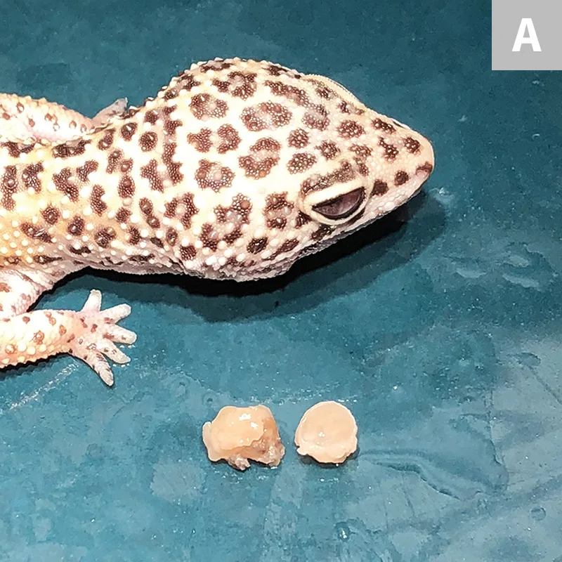
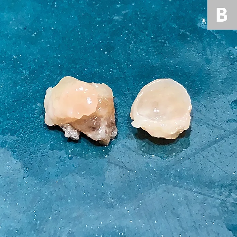
Leopard gecko from Figure 2 with solid cellular debris in both eyes due to vitamin A deficiency shortly after debris removal (A), with enhanced view of removed cellular debris (B)
Herbivores (eg, tortoises, green iguanas [Iguana iguana]) and omnivores (eg, bearded dragons [Pogona vitticeps]), along with carnivorous lizards and snakes that eat the entire body of an animal, generally are not affected by vitamin A deficiency. Omnivorous Emydidae turtles (eg, box turtles [Terrapene spp]) and red-eared sliders (Trachemys scripta elegans) can develop vitamin A deficiency.
Diagnosis
Diagnosis of vitamin A deficiency is based on dietary history and clinical signs. Because most vitamin A is stored in the liver and circulating levels do not fall until liver reserves are exhausted, circulating levels may not be an accurate indicator of overall vitamin A status. Normal values for liver or circulating levels of vitamin A or retinol are often unknown. Required blood and liver biopsy sample sizes may be greater than can be obtained safely in small patients, and incorrect sample handling and assay techniques can alter values.
Differential diagnoses for ocular and gingival disease may have numerous causes, including infectious (eg, bacterial, fungal, parasitic, viral), neoplastic, metabolic (eg, calcium deficiency [exposure gingivitis], uric acid deposits [conjunctivitis, retinal detachment, blepharospasm]), traumatic, and environmental (eg, from substrate, ultraviolet light, inappropriate humidity).
Fluorescein staining may reveal corneal ulcers. Corneal cytology may disclose secondary bacterial, fungal, parasitic, or neoplastic disease.
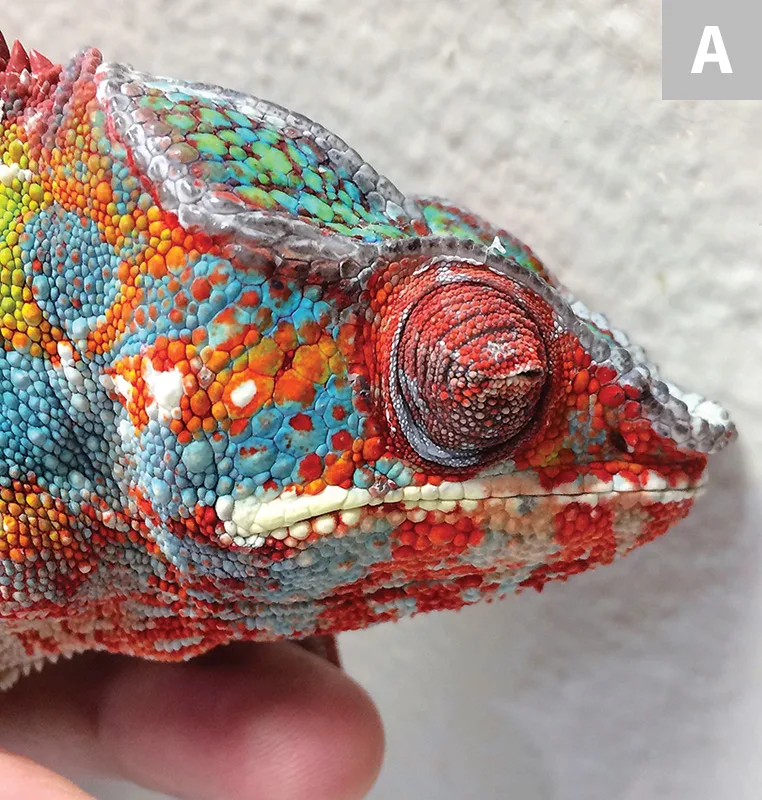
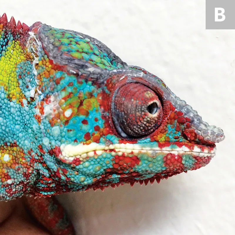
Panther chameleon with vitamin A deficiency and blepharospasm on presentation (A) and 2 weeks after a single injection of vitamin A palmitate (B). The eye is markedly improved, but some blepharedema remains.
Treatment
A vitamin A supplement should be added to the patient’s diet. All vitamin A doses are empiric and range from 5000-66 666 IU vitamin A palmitate per kg IM, SC, or PO every 1 to 2 weeks for 2 treatments.1,3 Injectable vitamin A has been associated with hypervitaminosis A; oral supplementation is preferred. Fat-soluble vitamin A is much less toxic than water-soluble vitamin A.
Broad-spectrum ophthalmic antibiotic ointments or antibiotic drops should be administered to lubricate and prevent secondary bacterial infection of the cornea, if compromised. In leopard geckos, solid cellular debris should be moisturized under the eyelids with saline and removed with blunt probes, hemostats, or fine forceps. The patient should be checked for ulcers using fluorescein staining, and eyes should be flushed copiously with saline. Similarly, eyes of chameleons should be flushed with sterile saline to remove thickened mucus and accumulated cellular debris secondary to xerophthalmia. Retained sheds around the eyes and feet and retained hemipenal casts should be removed with the patient under anesthesia.
If the patient has not eaten recently, nutritional support should be provided via fluid therapy and oral caloric supplementation. If the patient is eating, the quality of its diet should be improved. Owners should be instructed to gut load feeder insects with a diet containing vitamin A, at least 8% calcium, multivitamins, trace minerals, proteins, carbohydrates, and fat; to dust all insects with calcium before each patient feeding; and to feed multivitamins containing vitamin A—rather than calcium—twice a month.
Wild insectivorous reptiles eat hundreds of invertebrates (eg, insects, arachnids, mollusks, crustaceans), other lizards, mammals, and birds, so pet lizards should be fed as wide a variety of insects as possible; diet should not be restricted to crickets, mealworms, super or king mealworms, waxworms, and/or Dubia roaches. Reptile specialty stores and online vendors sell a wide variety of insects, including other cricket species (eg, black, field, banded), silkworms, black soldier fly larvae (sold as Phoenix worms), tobacco or tomato horn worms (sphinx or hawk moth larvae [sold as goliath worms or green giants]), butterworms, bean beetles, fruit flies, springtails, and wood lice, as well as wild-caught seasonally available insects (eg, moths, cicadas, flies, grasshoppers, katydids, bees [with stingers removed], cockroaches, crustaceans [eg, pill bugs, roly-poly bugs], mollusks [eg, snails, slugs]). Fireflies contain highly toxic lucibufagins and should never be fed to pet lizards.4 Some insectivorous reptiles will also eat neonatal mice, which are rich in vitamin A.
Patients should be re-evaluated every 1 to 2 weeks until they are eating readily and appear healthy. Body weight should be monitored to determine if nutritional supplementation is indicated, and dietary recommendations should be reviewed with the owners at each appointment to ensure they understand and have implemented the changes.
Precautions
In one study of cricket gut-loading diets, 3 of 4 diets did not improve the crickets’ calcium content because they were not calcium enriched.5 Feeder insects should be fed a gut-loading diet high in calcium. Calcium-fortified, high-moisture cricket wafers or high-moisture foods (eg, gel water cubes) are ineffective at increasing calcium or vitamin A content and are not recommended, even as a water source.6 Studies have shown that cubes decrease the effectiveness of gut-loading diets. Larvae will take water from damp paper towels, and other insects will drink from water-filled cotton balls in a bottle or syringe cap. Multivitamins should be discarded if no vitamin A is present, the supplement has expired, or the supplement is more than a year old.
Prognosis
Prognosis is poorer when clinical signs are more advanced or have been present for 6 months or longer. Patients with concurrent disease (eg, hepatic lipidosis, nutritional secondary hyperparathyroidism) also have a poor prognosis. If vitamin A deficiency is diagnosed early and treated appropriately, most patients will make a full recovery.
Patients with long-term vitamin A deficiency may be blind, and patients with severe cases may have corneal fibrosis similar to chronic canine keratoconjunctivitis (Figure 5). Patients with a history of anorexia lasting several months may have hepatic lipidosis and may be susceptible to refeeding syndrome.
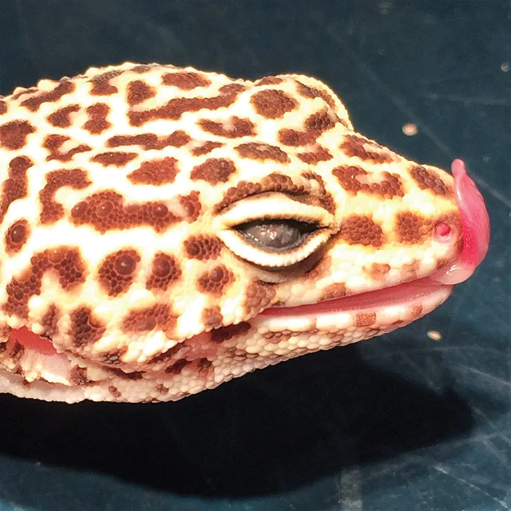
Corneal scarring in a leopard gecko as a sequela to chronic untreated vitamin A deficiency
Conclusion
Vitamin A deficiency in insectivorous reptiles is common and can result in severe epithelial disease, especially involving the eyes. Close attention to diet can prevent and diagnose this deficiency. Insectivores must be fed a varied diet, insects should be dusted with multivitamins containing vitamin A twice monthly, and, most importantly, feeder insects must be fed a balanced diet that contains vitamin A and 8% calcium. Clinicians should discuss these requirements in detail with all owners of insectivorous reptiles and amphibians.