Vertebral Fractures & Luxations in Dogs & Cats
Ashley Schenk, DVM, MPH-VPH, Lauderdale Veterinary Specialists, Fort Lauderdale, Florida
Adesola Odunayo, DVM, MS, DACVECC, University of Florida
Amy Hodshon, DVM, MS, DACVIM (Neurology), University of Tennessee
What do I need to know about vertebral fractures and luxations in dogs and cats?
Vertebral fractures and luxations most commonly occur secondary to trauma when excessive or unnatural forces (eg, motor vehicle accidents, falls, bite or gunshot wounds) are applied to the axis of the spine.1 Early recognition and stabilization of traumatic spinal cord injuries are imperative to positive patient outcome and return of normal neurologic function.
Clinical Signs
Patients are often presented shortly after a traumatic incident, most often with concurrent injuries. Some patients may show signs of systemic shock (eg, weakness, dyspnea, poor pulse quality, pale mucous membranes).2 Pulmonary contusions and further thoracic trauma can lead to dyspnea and poor oxygen saturation. Arrhythmias may result from myocardial hypoxia (associated with hemodynamic shock) as well as direct myocardial damage from blunt force.3 In addition, some patients may have long-bone or pelvic fractures and/or show signs of traumatic brain injury, including changes in level of consciousness, respiratory pattern abnormalities, decreased pupil responsiveness, and/or abnormal ocular movements and position.4
Diagnosis
It is crucial to perform a thorough physical examination and address airway, breathing, circulation, and dysfunction of other organ systems.1,5,6 Blood pressure stabilization is essential in maintaining spinal cord perfusion and allowing reliable assessment of mentation and nociception (ie, deep pain perception). Following systemic evaluation and stabilization of vital parameters, a full, careful neurologic examination should be performed.
Neurologic Examination & Localization
During the neurologic examination (Table), patient manipulation should be minimized until vertebral column instability can be ruled out. This can be accomplished by securing patients to a rigid “backboard” to provide temporary external coaptation and protect from further displacement.
Table: Initial Neurologic Examination7
*Testing the patient’s nociception is extremely important, as lack of nociception indicates a poor prognosis. This should be conducted in any limb in which voluntary movement is not seen to evaluate for further nerve injuries (eg, brachial plexus avulsion).
Sedatives are contraindicated for the initial examination, as they can interfere with neurologic assessment. They may also decrease the protective tone of the paravertebral muscles—which may decrease the stability of the vertebral column—and may adversely affect the cardiovascular system and further compromise systemic and spinal perfusion.5
After the initial neurologic examination has been performed and the patient has been immobilized, sedatives and analgesics may be administered cautiously for pain management and imaging.
Radiographs & Advanced Imaging
Survey radiographs of the vertebral column should be obtained, as well as chest and abdominal radiographs to assess for concurrent injuries. Horizontal beam radiographs are strongly preferred for obtaining ventrodorsal views of the vertebral column, as they do not require the patient to be moved out of lateral recumbency, which could further compromise the integrity of the vertebral column. Although sufficient for diagnosing significantly displaced vertebral fractures and luxations, radiographs are not adequate for predicting the degree of spinal cord injury or assessing the degree of vertebral instability (Figure 1 and Figure 2).
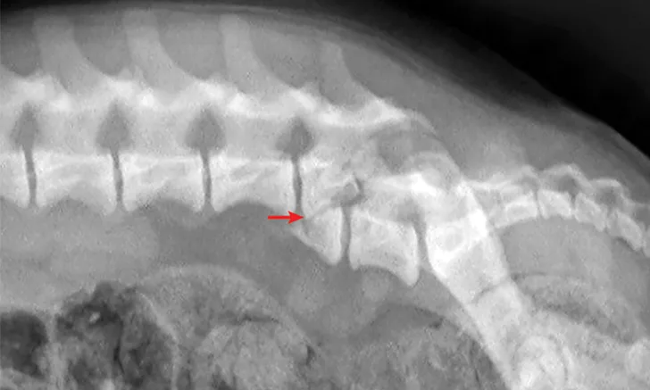
Figure 1
Lateral radiograph of a dog following unknown trauma. There is a complete oblique fracture (arrow) of the L6 vertebral body and luxation of the L6-L7 articular processes, with severe cranioventral displacement of the caudal segment. Despite the degree of displacement, this patient retained voluntary movement in both pelvic limbs on initial examination and made a functional recovery following reduction and stabilization of the fracture/luxation and 8 weeks of crate confinement.
Advanced imaging such as CT and/or MRI may be necessary to diagnose more subtle fractures or luxations, traumatic intervertebral disk herniations, or fracture fragments within the vertebral canal. Advanced imaging improves surgical planning by allowing visualization of potential spinal cord compression from hematoma, extruded disk material, or vertebral fragments; this helps the surgeon determine whether it is necessary to perform a hemilaminectomy in addition to vertebral stabilization. Advanced imaging before surgery also allows for better implant planning.7-9
Other Diagnostics
Additional diagnostics (ie, CBC, serum chemistry, urinalysis, arterial blood gas, thoracic and abdominal radiography, focused assessment with sonography for trauma) should be performed in patients with polytrauma to further characterize the involvement of other organ systems.10
Treatment
The patient’s overall condition, including signs of systemic shock and concurrent injuries, must be addressed. Comprehensive patient stabilization may include fluid therapy, analgesia, and antibiotics. Determining the appropriate treatment plan for the vertebral fracture or luxation depends on several factors: presence and nature of concurrent injuries, location of the spinal trauma, extent of displacement, severity of neurologic deficits, and owner goals and financial considerations.
Conservative management is generally preferred for fractures that have not caused vertebral instability and for many cervical fractures and/or luxations. It is also preferred for patients in which a correctly applied brace can effectively immobilize the injured segment and for those in which the risk for death with surgical stabilization is high.11 Patients with thoracolumbar or lumbosacral injuries should undergo advanced imaging and surgical treatment as soon as possible if spinal instability is noted on radiographs, if the neurologic deficits are significant (ie, weak or no voluntary movement seen), or if they worsen over time. Prognosis for functional recovery in patients with absent nociception resulting from vertebral fracture or luxation is poor, and euthanasia should be considered if a permanent cart or wheelchair is not an option.
Determining whether a vertebral injury has led to vertebral column instability can be difficult. Vertebral luxations are inherently unstable, but vertebral fractures may or may not cause instability, depending on which structures are affected. One model for assessing potential instability divides the vertebral column into 3 compartments (Figure 3); if 2 of the 3 compartments are disrupted, the injury has likely compromised vertebral column stability, and additional corrective measures should be taken.
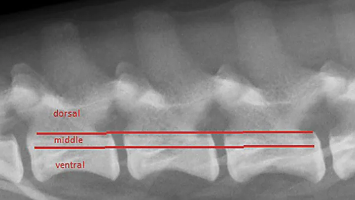
Three-compartment model of the vertebral column. The dorsal compartment includes structures dorsal to the line (ie, pedicles, laminae, articular processes, interarcuate ligaments, spinous processes). The middle compartment includes the dorsal aspect of the vertebral body, the intervertebral disk, and the dorsal longitudinal ligament. The ventral compartment includes structures ventral to the line (ie, the remainder of the vertebral body and intervertebral disk and the ventral longitudinal ligament).
Medical Management & External Coaptation
Conservative management of vertebral fractures and vertebral trauma should always include appropriate analgesia and strict cage rest with or without external coaptation devices such as spinal braces. External coaptation devices can protect against dorsoventral angulation of the thoracolumbar spine but require significant monitoring and nursing care.5,12,13 The cervical vertebrae are typically the easiest to immobilize with a brace (Figure 4). Conservative management may be more difficult and less successful in feline patients because their spinal injuries are often more severe and because cats do not tolerate splints and cage rest as well as do most dogs.
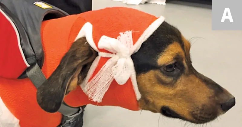
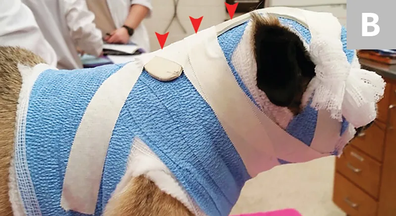
(A) Neck braces must extend from in front of the patient’s ears to behind the thoracic limbs to adequately immobilize the cervical spine. (B) A molded, rigid component (arrows) is usually incorporated either dorsally or ventrally into the bandage. Clients should be instructed to monitor for any difficulty breathing, chewing, or swallowing, and the bandage must be changed every 2 to 3 weeks to prevent dermatologic complications. B courtesy of Rebecca Hodshon, DVM, DACVS
Surgical Decompression & Stabilization
Surgical decompression and stabilization are often recommended for thoracic and lumbar injuries based on the degree of instability and risk for further vertebral column shifting and spinal cord damage. Surgical options include use of positive profile pins or screws with polymethylmethacrylate bone cement, external fixators, and vertebral body plates (Figure 5). Referral to a board-certified neurologist and/or surgeon is recommended for cases in which surgical therapy may be an option. Cats with sacrocaudal luxations may benefit from tail amputation, which removes a source of pain and prevents further traction damage to the sacral nerve roots.
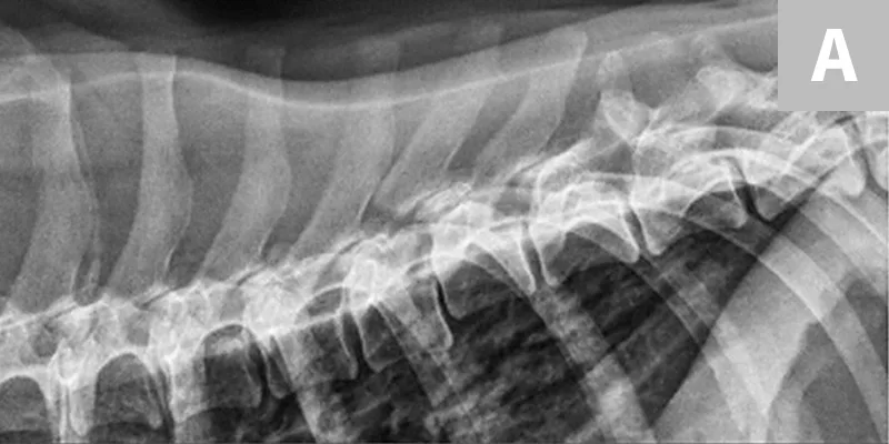
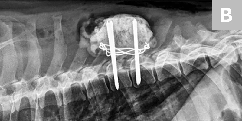
Lateral radiographs taken pre- and postoperatively of a dog after being found in a ditch and presumably hit by a car. (A) There is a T8-T9 subluxation and (inconsequential) T8 spinous process fracture. (B) The subluxation was reduced and stabilized using fully threaded pins, Kirschner wire, and bone cement. The Kirschner wire encircles the pins and is intended to increase the strength of the bone cement. Ideally, these pins would have been placed with more craniocaudal angulation and the construct would have included a third implant spanning the luxation laterally to provide additional support.
Steroid Use
Use of glucocorticoids in managing spinal cord injuries remains controversial. Methylprednisolone sodium succinate has free radical scavenging effects at high doses and has been shown to have some efficacy in humans with spinal cord injury when treatment is initiated within 3 to 8 hours of the initial injury; however, the observed degree of improvement was so slight that it was considered irrelevant in veterinary patients.14 A recent randomized controlled study evaluating dogs with severe spinal cord trauma caused by intervertebral disk herniation found no benefit to administering methylprednisolone sodium succinate.15 Given the lack of proven benefit and the risk for adverse events, use of steroids is not recommended.13,16
Euthanasia
Euthanasia should be considered in patients with absent nociception caused by a spinal fracture or luxation or when euthanasia is in accordance with the owners’ goals, expectations, and financial limitations. In cases of lumbosacral or sacrocaudal injuries, addressing the potential for permanent urinary and/or fecal incontinence is an important part of communicating possible outcomes to owners.5
Prognosis
The prognosis for vertebral injuries is directly related to the degree of the associated spinal cord injury. The most important neurologic deficit to recognize is the absence of nociception caudal to the suspected lesion. This is seen almost exclusively in injuries to the thoracolumbar and lumbosacral spinal cord. Patients without nociception resulting from a vertebral fracture or luxation have a grave prognosis for return of function.12 Conversely, if the patient sustains a thoracolumbar injury and retains nociception, the prognosis for return of function is considered good (≈80% in dogs) as long as rapid decompression and/or stabilization are obtained.17 Although the presence of Schiff-Sherrington posture can help localize the injury to the thoracolumbar spinal cord, it carries little to no prognostic value.
The prognosis for cervical injuries is usually good, even with nonsurgical management, as long as the patient retains some voluntary motor function.5,7 The prognosis for sacral fractures is also generally good18,19; however, persistent pain due to nerve root entrapment and potentially permanent urinary and fecal incontinence can be reasons for euthanasia. Cats with sacrocaudal injuries that retain tail-base sensation have a good prognosis for recovery of urinary continence.20
Conclusion
Although uncommon, vertebral fractures, luxations, and subluxations are important to consider when managing patients with trauma. Early recognition and stabilization decisions are the basis for ensuring the best chance for recovery. Appropriate treatment decisions must be made in consideration of neurologic deficits and the owner’s long-term goals. With medical and surgical management, treatment options must be tailored to the individual case. Appropriate nursing care, adequate exercise restriction, and a slow, gradual return to activity are crucial in the long-term recovery of spinal trauma patients.