Urine Crystals in Dogs & Cats
Microscopic examination of urine sediment should be included as part of a complete urinalysis. Crystalluria is not typically of clinical significance and is often not associated with urolithiasis; however, the presence of abnormal crystals or large numbers of common crystals may indicate disease.
Metabolism, diet, underlying disease, sample collection, sample storage, and iatrogenic causes (eg, drug or IV contrast agent administration) may contribute to crystalluria. Crystal formation is often dependent on urine pH, osmolality, and temperature, as well as concentration and solubility of the crystal-forming material. Identification of urine crystals is typically based on their shape and color.

Figure 1A
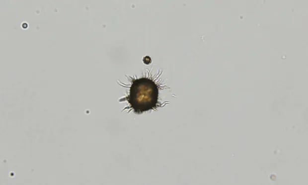
Ammonium biurate crystals appear as yellow to brown spherules with irregular projections (“thorn apple” or “sarcoptic mange” appearance). A smooth, spheroid form may also be seen. Crystals typically form as a result of liver disease or portal vascular anomalies. Decreased conversion of ammonia to urea results in hyperammonemia, and increased concentration of ammonia in the urine leads to crystal formation. These crystals may also be seen in animals with ammonium biurate urolithiasis. In general, they should be considered a pathologic crystal indicative of underlying hepatic disease; however, they may be seen in low numbers in clinically normal dalmatians and English bulldogs.