Treatment of Melting Corneal Ulcers in Dogs
Georgina M. Newbold, DVM, DACVO, The Ohio State University
In the literature
Guyonnet A, Desquilbet L, Faure J, Bourguet A, Donzel E, Chahory S. Outcome of medical therapy for keratomalacia in dogs. J Small Anim Pract. 2020;61(4):253-258.
The Research …
Keratomalacia (ie, corneal melting) can be a vision-threatening problem in dogs. These challenging cases often are not presented to the clinician until there is already significant corneal stroma lost to collagenolysis (ie, rapid tissue destruction by collagenase and protease enzymes).1-4 These enzymes are a constitutive part of the normal corneal environment and help balance corneal health; however, they can become increased in cases of bacterial infection.1,2 A recent study examining dogs with melting corneal ulcers found that several predisposing factors were present in these patients4; the study also described the clinical outcome, including vision, corneal scarring, and need for corneal surgery, in dogs with keratomalacia.4
Brachycephalic dogs (eg, French bulldogs, pugs, shih-tzus), dogs with pre-existing corneal disease (eg, keratoconjunctivitis sicca), and dogs with systemic problems (eg, diabetes mellitus) may be predisposed to keratomalacia.4 Once diagnosed, keratomalacia should be treated aggressively, usually with multiple topical medications at very frequent administration intervals. Corneal cytology and culture with susceptibility testing can help guide medical therapy. As bacterial resistance and mixed-bacterial infections become more common, antibiotic monotherapy may not be the best approach for these patients. In addition, use of collagenase inhibitors to halt tissue destruction is key to preventing progression of corneal damage.
In this retrospective study, 57 melting ulcers in 53 dogs were treated with ophthalmologic tobramycin (every 2-4 hours) and equine serum (every 2-4 hours). Of these ulcers, 31 out of 57 (54.4%) healed within 15 days (median, 5 days) with medical therapy alone. Significant progression of keratomalacia necessitating surgical intervention occurred in the remaining 26 eyes. This study also found that 16 out of 27 (59.2%) bacterial isolates were resistant to tobramycin and other topical antibiotics.4 Frequent application of equine serum potentially helped to diminish collagenolysis; however, the use of additional protease inhibitors may have improved overall results. Although the long-term visual outcome was satisfactory in almost all cases, 26 out of 57 (45.6%) ulcers ultimately required surgery to treat keratomalacia.
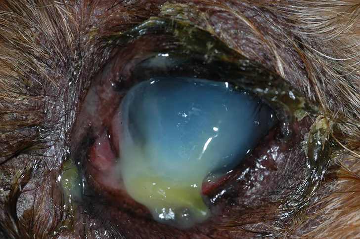
Keratomalacia in a Chihuahua with dry eye
… The Takeaways
Key pearls to put into practice:
Corneal cytology and culture and susceptibility testing is recommended in all dogs with deep or melting ulcers. In-clinic cytology can help direct antibiotic therapy, as culture results may take several days.
Multimodal antibiotic therapy (eg, combinations of topical antibiotics with both gram-positive and gram-negative activity) may help improve the spectrum of coverage. Potential combinations include tobramycin/cefazolin, tobramycin/chloramphenicol, and ofloxacin/cefazolin.
Multimodal anticollagenase therapy, such as combined use of topical serum (canine or equine), topical K+EDTA, and oral doxycycline, may help slow progression of keratomalacia.
Frequent, rigorous (eg, every 2 hours during waking hours, every 4 hours overnight) application of topical medication is important in medical management of melting ulcers. Frequent (every 1-2 days) recheck examinations are also important to determine if surgical stabilization is necessary. Referral to a veterinary ophthalmologist early in the course of disease may improve long-term outcome.