Top 5 Viral Dermatoses in Cats
Liora Waldman, BVM&S, CertSAD, MRCVS, Veterinary Dermatology & Allergy Center, Haifa, Israel
Alexander Werner Resnick, VMD, DACVD, Animal Dermatology Center
Hyperkeratotic hyperpigmented plaque on the nose, muzzle, and chin of a cat infected with papillomavirus
Viral dermatoses in cats are rare diseases caused by direct viral cytopathic effects in the skin. Viral infections are diagnosed using techniques such as PCR, immunohistochemistry, and in situ hybridization. Presented below are the authors’ top 5 feline viral dermatoses most likely to be encountered in veterinary practice.
1. Papillomavirus
Papillomavirus influences cell growth and differentiation and may cause cancer.1 Although in general the viruses are species-specific, human and bovine papillomaviruses have been detected in cats.2 Four feline papillomaviruses have been completely sequenced. FcaPV-2 is the most frequently isolated from cat lesions1,3,4 and was found on the skin of 52% of cats in one study.5 FcaPV-2 and FdPV-3 are closely related to canine PV-1 and canine PV-7,3 respectively.
Papillomavirus causes the following dermatoses in cats:
Viral plaques are uncommon and are seen as single or grouped round-to-oval scaly, gray, tan, or black papules or plaques with hyperkeratosis (Figure 1).6 They are neither pruritic nor painful and may be present anywhere on the body. In healthy cats, they may resolve spontaneously.2 In immunosuppressed cats (eg, those with FIV, FIP, FeLV, or neoplasia; those receiving glucocorticoid treatment), resolution occurs after treating the primary cause. Demodex cati mites might be found in the lesions.1,6-9

Hyperkeratotic hyperpigmented plaque on the nose, muzzle, and chin of a cat infected with papillomavirus
Bowenoid in situ carcinoma (BISC; ie, Bowen’s disease) is often a progression from viral plaque. It presents as hyperpigmented macules or crusted plaques that may ulcerate. The face, neck, and limbs are predisposed, but lesions can be seen anywhere on the body.3 Cutaneous horns may be present.10 BISC can progress to squamous cell carcinoma with metastases.11 Ultraviolet light does not affect neoplastic transformation (Figure 2).2,8,9

Ulceration, bleeding, alopecia, and hyperkeratotic plaques on the face of a cat with Bowen’s disease caused by papillomavirus infection
Cutaneous papilloma is rare and appears as a single pedunculated or cauliflower-like hyperkeratotic lesion, smaller than 0.5 cm, at any body site.2,8
Oral papilloma is also rare. Only 2 cases have been reported, found on the tongue as small, multifocal, soft, pink, raised, flat-topped lesions.12
Feline sarcoid is seen in cats in rural areas and is likely caused by bovine papillomavirus.13 Sarcoid lesions are firm, round, single masses that can ulcerate and occur mainly on the nose, upper lip, and digits. They do not metastasize but may recur after excision.8-10
Basal cell tumors are uncommon and usually appear as a soft, mobile, painless, dermal mass with some surface scaling. A novel papillomavirus similar to FcaPV-314 has been identified.
Papillary squamous cell carcinoma is seen on the head as hornlike crusted masses.6
Fibrosarcoma and apocrine gland cysts are rarely associated with papillomavirus.2,15
Diagnosis is made via histopathology, PCR (swab more sensitive than formalin fixed tissue1), immunohistochemistry, in situ hybridization, or electron microscopy.9,10
Treatment of oral papilloma and single plaques may include excision, cryosurgery, electrosurgery, or CO2 laser ablation. Some plaques might regress following control of the primary problem. Imiquimod can be effective for plaques and BISC within 3 to 4 weeks. Lesions can recur when treatment is discontinued.8,10 Side effects may include local erythema and, occasionally, systemic effects (eg, nausea, vomiting, myalgia, fever, hypotension).16 Interferon-α (IFNα; 30 units PO q24h) has been reported to be effective.10
2. Feline Herpesvirus 1
FHV-1 primarily causes facial dermatitis affecting the nasal planum, muzzle, bridge of nose, and periocular skin (Figure 3), but it can occur at other sites.17-19 Recent upper respiratory infection, stress, and/or glucocorticoid therapy may precede onset.

Twelve-year-old domestic shorthair cat with FHV-1 dermatitis
Facial lesions may start unilaterally with vesicles, erythema, and alopecia. Due to intense pruritus, lesions may become ulcerated and crusted.
Differential diagnoses include allergy (eg, food, atopy) and eosinophilic plaque.19 Diagnosis is made via histopathology, revealing eosinophils, neutrophils or lymphoplasmacytic dermatitis, necrosis, or ulceration with intranuclear inclusion bodies. PCR from fresh biopsy (preferably in saline, not formalin) should be submitted for definitive diagnosis.19,20
Treatment options include famciclovir (90-125 mg/kg q8-12h19,20), recombinant feline interferon ω (1.5 million units/kg perilesionally and SC for 2-3 weeks) or recombinant feline interferon-α (1 million units/m2 SC 3 times per week).21 Topical idoxuridine, cidofovir, or trifluridine is used to treat ulcerative keratitis. Vaccination can protect cats from developing lesions.
3. Feline Calicivirus
Feline calicivirus is an RNA virus that is shed via ocular, nasal, and oral secretions.10,22
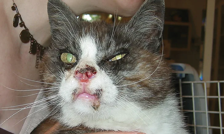
Dermatologic signs may include ulcers on the nasal philtrum, lips, tongue, gingiva, and paws10; swollen feet; facial skin erosions, especially of the nose; and ventral pustules (Figure 4).21

Ulceration with crusting on the nose, philtrum, muzzle, and upper lip of a calicivirus-infected cat. Image courtesy of Stephen White, DVM
Differential diagnoses include nasal ulceration (eg, from FHV-1, squamous cell carcinoma, other neoplasia, Cryptococcus spp, Sporothrix schenckii, or mosquito hypersensitivity), and ulceration of the paws (eg, from pox virus, papillomavirus, FeLV, malignancy, plasma cell pododermatitis).
Treatment with antibiotics might be needed if secondary bacterial infection develops. Oral glucocorticoids may be beneficial to treat oral ulceration.
4. Feline Pox Virus
Feline pox virus dermatitis, caused by cowpox virus, is a rare disease seen primarily in Europe and West Asia. Cats are infected by hunting rodents (the natural hosts), typically in rural areas. Lesions occur mostly on the head, ears, neck, and legs. Primary lesions may look like bite wounds, nodules, plaques, crusted papules, ulcers, abscesses, or cellulitis (Figures 5 and 6). Pruritus is variable. Crust-covered, ulcerated papules and nodules typically develop within 1 to 3 weeks. Oral ulceration can lead to anorexia. Fever, conjunctivitis, and pneumonia may develop.10,23,24

FIGURE 5
Ulceration on the head and face and crusted ulcer in the medial canthus of the left eye in a cat infected by cowpox virus. Image courtesy of Tim Nuttall, BVSc, PhD
Pox virus is zoonotic, especially in immunosuppressed humans.
Differential diagnoses include bacterial and fungal infections, neoplasia (mast cell tumor, lymphoma), and granuloma. Diagnosis is made via biopsy (eosinophilic intracytoplasmic inclusion bodies), serology, PCR, immunohistochemistry, or virus isolation.
Most patients recover without complications. Treatment may include antibacterial drugs for secondary infections and supportive treatment. Glucocorticoids are contraindicated.
5. Feline Leukemia Virus
FeLV, a retrovirus, causes giant-cell dermatosis10,25 with pruritus, ulceration, and crusting lesions—mainly on the head, neck, and face (Figure 7) but occasionally on the extremities or footpads, trunk, and mucocutaneous junctions of the anus and/or prepuce. Cutaneous horns may be seen.10

Thirteen-year-old FeLV-positive cat with alopecia, mildly seborrheic dermatitis, and excoriations on the ventral neck
Differential diagnoses include evident pruritus caused by allergy (eg, food, atopy), Notoedres cati, Cheyletiella spp, or Demodex spp; or crusted lesions caused by exfoliating dermatitis, pemphigus foliaceus, drug reaction, systemic lupus erythematosus, or seborrhea. Diagnosis is made via histopathology (giant-cell dermatoses), serology, or PCR.10,25,26