Top 5 Tips for a Successful Dermatological Examination
Jackie Davis, LVMT, University of Tennessee
Skin and ear problems are 2 of a veterinary practice’s most common presenting complaints. Dermatologic diseases, which can be mild or severe, exhibit many different signs (eg, skin lesions, pruritus, ear disease symptoms). A successful dermatologic examination is essential for ensuring a correct diagnosis and prognosis.
1. Take Time for a Thorough History
A complete history is essential for making a correct diagnosis, and seemingly minor details can be important clues to the underlying cause. Ask clients to complete a questionnaire designed specifically for dermatologic patients to be reviewed with the veterinary technician before the physical examination. The more detailed the history, the easier it will be to make a diagnosis and treatment plan. It is important to include specific questions such as:
Does the patient lick, scratch, or chew?
How itchy is the patient? (Ask the client to grade on a scale of 0–10, with 0 indicating no evidence of pruritis and 10 indicating constant scratching, licking, and chewing.)
Where does the patient itch?
Does the patient itch only in certain seasons?
When did the problem start?
What is the patient’s environment (eg, indoor or outdoor)?
2. Look Everywhere, Feel Everywhere
Some lesions that are small or covered by hair are easily missed, so skin palpation is necessary.
Good lighting in the examination area is essential. Start the patient examination at the tip of the nose and work to the tip of the tail. Spread skin folds and toes apart to expose any hidden lesions (Figure 1). Part the hair, because some lesions may not be easily palpable or may be nonpalpable.

The patient's interdigital erythema could not be visualized until the toes were spread apart.
Roll the patient laterally or dorsally to better visualize the abdominal and axillary areas. Examine the ear canals and tympanic membranes with a handheld otoscope.
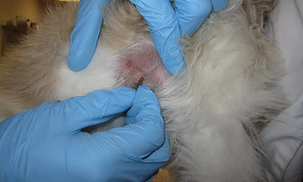
3. Always Perform Diagnostics
Simple diagnostics can eliminate many conditions and are easy and quick to perform and interpret.
Skin scraping: Rules out demodicosis (Figure 2)
Skin cytology: Determines the presence of bacteria, yeast, or inflammatory cells (Figure 3)
Ear cytology: Differentiates between a bacterial or yeast infection
Fine-needle aspirate: Helps diagnose possible neoplasia
Trichograms: Useful for examining the structure of the hair shaft, tip, and bulb; look for broken hairs, pigmentation abnormalities, fungal spores, and Demodex.

When performing a deep skin scrape, obtaining a small amount of capillary blood is necessary. Demodex canis lives in the hair follicles and can be missed if the scraping is not deep enough.

If moist exudate is on the skin, take an impression smear by pressing a glass slide onto the exudate to obtain a cytology sample. If the lesion is dry, use clear cellophane tape to obtain a sample of the surface skin cells.
4. Develop Excellent Microscope Skills
The team’s ability to interpret scrapings and cytologies in-house makes the visit more efficient and less costly (Figure 4). Invest in a high-quality microscope and cytology and/or parasitology books for reference. Consider having sample slides with various organisms, external parasites, and normal and abnormal cell types for reference. Practice and improve microscope skills to boost experience and confidence.

An impression smear is stained using a modified Wright-Giemsa stain. Ideally, stain used for skin and ear cytologies should not be used for blood smears and should be changed more frequently to prevent cross-contamination.
5. Plan Extra Time for Client Education
Client education is an important part of the dermatologic visit. Clients must understand the disease process, be able to administer the recommended treatments, and be aware that they are integral to the treatment plan’s success. Educate clients in the examination room and at discharge, and support and encourage them throughout home treatment. This may be overwhelming for clients, so provide educational handouts they can review and refer to at home.
Conclusion
The veterinary technician should assemble the history, physical examination findings, and diagnostic results for the veterinarian to diagnose the primary disease and any secondary infections. The veterinarian should then develop a treatment plan based on the diagnosis and any client concerns.
This article originally appeared in the June 2015 issue of Veterinary Team Brief.