This response is incorrect. See below for the correct answer.
Top 5 Radiographic Variants
Anthony Pease, DVM, MS, DACVR, Viticus Group

Interpreting radiographs can be difficult when a lesion is not obvious. Clinicians must consider anatomic differences among species and breeds to understand what to expect on radiographs and how to differentiate normal from abnormal radiographs.
Both dog size and breed may affect the appearance of the thorax. For example, clinicians should not expect the thorax of a Chihuahua and a greyhound to display the same radiographic appearance. Similarly, a cat should not be compared with a small dog, as geriatric changes to the heart and appearance of the abdomen, including renal size, are not standard between cats and dogs.
Following are the author’s top 5 canine and feline radiographic variants.
1. Cardiac Silhouette of Cats on Thoracic Radiographs
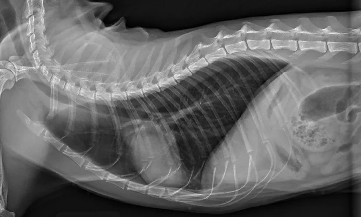
Thoracic radiographs are generally difficult to assess in cats due to age-associated variability. The cardiac silhouette develops increased sternal contact as cats age (Figure 1), with the aorta creating a notch or right-angled appearance that can falsely suggest left atrial enlargement on ventrodorsal projection. The aortic arch may have a rounded appearance on the ventrodorsal projection that can be mistaken for a pulmonary nodule in the left cranial lung lobe (Figure 2).1
Trusted content.
Tailored to you.
For free.
Create an account for free.
Want free access to the #1 publication for diagnostic and treatment information? Create a free account to read full articles and access web-exclusive content on cliniciansbrief.com.