Top 5 Radiographic Variants
Anthony Pease, DVM, MS, DACVR, Viticus Group

Interpreting radiographs can be difficult when a lesion is not obvious. Clinicians must consider anatomic differences among species and breeds to understand what to expect on radiographs and how to differentiate normal from abnormal radiographs.
Both dog size and breed may affect the appearance of the thorax. For example, clinicians should not expect the thorax of a Chihuahua and a greyhound to display the same radiographic appearance. Similarly, a cat should not be compared with a small dog, as geriatric changes to the heart and appearance of the abdomen, including renal size, are not standard between cats and dogs.
Following are the author’s top 5 canine and feline radiographic variants.
1. Cardiac Silhouette of Cats on Thoracic Radiographs
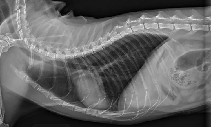
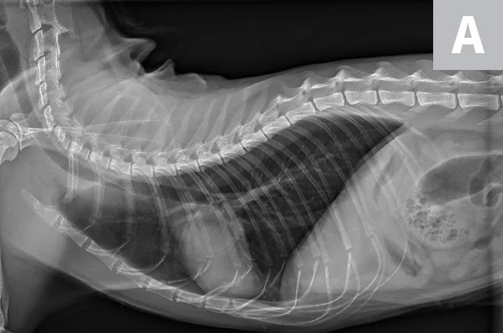
Thoracic radiographs are generally difficult to assess in cats due to age-associated variability. The cardiac silhouette develops increased sternal contact as cats age (Figure 1), with the aorta creating a notch or right-angled appearance that can falsely suggest left atrial enlargement on ventrodorsal projection. The aortic arch may have a rounded appearance on the ventrodorsal projection that can be mistaken for a pulmonary nodule in the left cranial lung lobe (Figure 2).1

FIGURE 1A
Lateral radiographs of a 2-year-old cat (A) and a 9-year-old cat with increased sternal contact of the cardiac silhouette (B).
2. Cardiac Silhouette of Dogs on Thoracic Radiographs
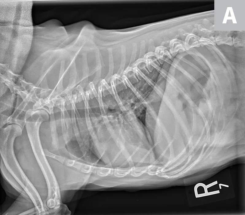
The cardiac silhouette can appear larger in small-breed dogs because the heart occupies a large amount of thoracic space; conversely, the cardiac silhouette can appear smaller in large-breed dogs (eg, greyhounds) due to the relatively larger size of the thorax (Figure 3).2
In dogs and cats, the vertebral heart score (VHS) system measures the width of the cardiac silhouette (ie, distance from cranial to caudal [ie, short axis] along the estimation of where the atria and ventricles meet) and from the carina of the trachea to the apex of the heart at its most ventral point (lines [ie, long axis] should be perpendicular to each other). These measurements are transferred caudally starting at T4 to calculate the VHS (Figure 4).2 Although this measurement is a good general guide, it can be overinterpreted, as cardiac chambers can change size without changing the shape of the cardiac silhouette.3

FIGURE 3A
Relative heart size difference on lateral thoracic radiographs of a normal basset hound (A) and a normal greyhound (B).
The average VHS in dogs is approximately 9.5 ± 0.5 with a normal range of 8.7 to 10.7. The measurement can change based on right or left recumbency (see a compilation of breed-specific VHS reference ranges available from Nguyenba in Suggested Reading). Measurements are more useful when diagnosing dilatative forms of cardiac disease and are reported to be less accurate in dogs with cardiac diseases with concentric hypertrophy.3 VHS for cats in right lateral recumbency is 7.3 ± 0.5.4 A VHS >7.9 has a high diagnostic accuracy in distinguishing cats with left-sided cardiac disorders from healthy cats.5
Because there are breed differences in VHS measurements, finding a reference range is valuable and can increase accuracy of the assessment.3,6 In a study, VHS measurements were found to be most accurate in Yorkshire terriers and Cavalier King Charles spaniels.3 Heart size relative to thoracic volume varies among dog breeds and is considered to be primarily due to thorax conformation. In barrel-chested breeds (eg, bulldogs, Boston terriers, Lhasa apsos), the cardiac:thoracic ratio is generally larger than in normal or deep-chested breeds. The VHS system was developed to help address this issue but its efficacy is unclear (see Benefits & Limitations of Vertebral Heart Scoring).7
Benefits & Limitations of Vertebral Heart Scoring
The benefit of VHS is limited because of the wide range of normal values. Normal variable VHS values have been determined for several breeds of dogs and cats. One of the greatest benefits of VHS, however, is that it allows for the comparison of recheck radiographs to monitor disease progression and response to treatment. Some studies have suggested that subjective heart assessment is better than VHS at differentiating cardiac disease.3,7 Studies have also suggested that interobserver variability of VHS can play an important factor in assessment of heart size.10
3. Kidney Placement & Size in Dogs on Abdominal Radiographs
Abdominal radiographs have less variability among dog breeds regarding overall appearance and location of organs. Kidney length has been reported to be 2.98 ± 0.44 times the length of L2 on the ventrodorsal view.8 Still, there are significant differences in renal:L2 size ratios between brachycephalic and dolichocephalic dogs. Small-breed dogs (<22 lb [10 kg]) have been shown to have a significantly higher left kidney:L2 ratio as compared with large-breed dogs (>66 lb [30 kg]). A single normal ratio should not be used for all dogs.
4. Kidney Size in Cats on Abdominal Radiographs
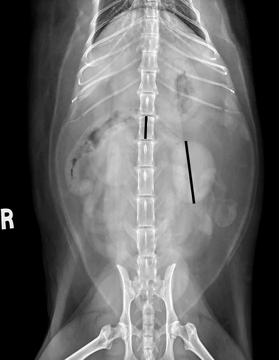
Kidney size varies between intact and neutered male cats.9 On ventrodorsal radiographs, normal kidney:L2 ratios are 1.9 to 2.6 (measuring renal length and dividing by the length of L2) in neutered male cats and 2.1 to 3.2 in intact cats (Figure 5).9

FIGURE 5
Ventrodorsal radiograph measuring renal size of normal kidneys in a neutered cat. The longer black line over the renal silhouette shows the length of the left kidney, which can be compared with the shorter black line superimposed on the L2 vertebral body.
5. Musculoskeletal Variations in Chondrodystrophic & Large-Breed Dogs
Chondrodystrophic musculoskeletal variations on radiographs are generally based on the difficulty of obtaining straight lateral and craniocaudal radiographs of the thoracic limbs (Figure 6) in chondrodystrophic dogs, whereas large-breed musculoskeletal variations on radiographs are generally based on the difficulty of obtaining straight lateral radiographs of the stifles and tarsi in large-breed dogs.
Positioning chondrodystrophic dogs for musculoskeletal radiographs can be challenging because of their curved humerus, radius, and ulna. It is generally easier to position the thoracic limbs of large-breed dogs as compared with chondrodystrophic dogs, but the pelvic limbs (primarily the tarsus and stifle) may appear oblique due to the large thigh musculature on lateral projections and the inability to completely extend the coxofemoral joints. Use of pads or passive restraint (eg, ropes, tape) and/or tilting the radiographic tube can help optimize the radiographic image. Literature focused on positioning and minimizing the difficulty in interpreting normal radiographs can be found in Suggested Reading.
Conclusion
Radiographs provide a quick overview of the thorax, abdomen, and musculoskeletal structures, but the interpretation can be variable. VHS measurement is a helpful guide for identifying and monitoring heart size changes but is not as accurate as echocardiography to determine the internal architecture and function of the heart. Age changes, breed variation, and neutered versus intact status can be associated with radiographic variability. Interpretation of abnormalities should be aided by various measurement parameters but always combined with clinical signs and physical examination findings.