Mydriasis and altered tapetal reflection in a cat secondary to serous retinal detachment
Hypertension is a common cause of vision changes and loss and increased risk for damage to the brain, kidneys, and heart.1-4 Ocular lesions are often an initial clinical sign (see Diagnosing & Treating Hypertension).
Following are the 5 most common ophthalmic sequelae of systemic hypertension in the author’s experience.
1. Decreased Vision/Blindness
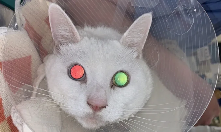
Decreased or altered vision, vision disturbance, and sudden blindness are emergencies. Patients with systemic hypertension may exhibit initial clinical signs of acute vision loss with varying degrees of mydriasis (Figures 1 and 2), negative or incomplete pupillary light reflexes, negative to varying degrees of light perception, and/or reduced ability to navigate a maze test or recognize humans.5 Systemic hypertension can cause visual disturbance or acute blindness from intraocular hemorrhage, subretinal edema, retinal detachment, or secondary glaucoma.1,6,7

Fixed, dilated pupil (common secondary to systemic hypertension) and subsequent serous retinal detachment in a cat
Differential diagnoses for changes in vision are old age, corneal disease, uveitis, cataracts, glaucoma, progressive retinal atrophy (dogs; less common in cats), retinal detachment, retinal edema, sudden acquired retinal degeneration (dogs), and optic nerve or intracranial disease.5 Common causes of vision loss in cats are uveitis, corneal disease, cataracts, glaucoma, retinal degeneration, retinal detachment, retinal edema, and optic nerve and intracranial disease.
2. Retinal Hemorrhage
Retinal hemorrhage is a common sequela to hypertension.1 Hemorrhages may be focal, multifocal, or large hemorrhagic subretinal areas (Figures 3 and 4). The mechanism is related to arteriolar vascular permeability changes.8,9 Autoregulatory mechanisms cause initial vasoconstriction, followed by possible arteriole lumen occlusion and ischemic necrosis. Increased vascular permeability can affect the choroid and cause subretinal fluid and retinal detachment.8,9

Fresh, characteristic keel-boat–shaped preretinal hemorrhages (arrows) between the retina and vitreous humor secondary to systemic hypertension in a dog

Multifocal retinal hemorrhages of varying shapes in the tapetum secondary to systemic hypertension in a dog
Retinal hemorrhages are also common with other conditions (eg, trauma, infectious disease, coagulopathy, neoplasia, diabetes, congenital disorders) that cause vasculitis, as well as hyperviscosity syndrome.8 In dogs, tick-borne diseases are infectious agents that can be associated with retinal hemorrhage.8
Differential diagnoses for retinal hemorrhage are coagulopathy, infectious disease, neoplasia, trauma, congenital disorders, and hyperviscosity syndrome secondary to multiple myeloma.
3. Hyphema/Intraocular Hemorrhage
Hyphema is a classification of intraocular hemorrhage that refers to a collection of erythrocytes in the anterior chamber (Figures 5 and 6). Intraocular hemorrhage can develop with any systemic condition that affects the vasculature of intraocular structures and can occur in the anterior chamber, surface of the iris, vitreous cavity, retina, choroid, supra- or subchoroidal space, and around or on the optic disc. Blood accumulates in the anterior chamber or vitreous humor due to disruption of the iris or ciliary body vessels in response to sustained systemic hypertension. Altered permeability of the uveal vasculature (iris, ciliary body, choroid) may then lead to intraocular hemorrhage,8,9 which may be visualized as a few strands of blood in the anterior chamber or on the iris surface, partial or complete hyphema in the anterior chamber, focal or large areas of vitreal hemorrhage, or retinal hemorrhage. Hemorrhage may be substantial and obscure large areas of the fundus, depending on chronicity and severity of systemic hypertension.

Ventral anterior chamber hyphema secondary to hypertension in a dog

Progressing hyphema secondary to hypertension in a dog that obscures intraocular structures
Differential diagnoses for intraocular hemorrhage are trauma, intraocular tumor, systemic neoplasia, coagulopathy, chronic glaucoma, chronic uveitis, and retinal detachment.10
4. Retinal Detachment
Exudative or serous retinal detachment is common in patients with hypertension.11 Clear to yellow fluid accumulation or blood in the subretinal space that may appear as the retina billowing toward the examiner is characteristic during ophthalmoscopy (Figures 7 and 8). Retinal detachment can also be secondary to vascular, inflammatory, and/or neoplastic diseases of the retina, choroid, and retinal pigmented epithelium.12

Serous retinal detachment in a cat with hypertension. Characteristic billowing retina (arrow) due to subretinal fluid is visible.

Characteristic subretinal edema and billowing of the retina (arrows) associated with systemic hypertension in a cat
Leaking fluid can overwhelm the retinal pigmented epithelium pumping mechanism, causing fluid accumulation in the subretinal space and leading to retinal detachment and vision loss. Vision loss may be acute or gradual and partial or complete, depending on the degree of vitreal hemorrhage and retinal edema. Correction of the underlying cause may allow resorption of subretinal fluid; vision recovery is possible if the condition is treated before retinal ischemia occurs. Morphology of the retina and retinal pigmented epithelium interface may not recover, even after 6 months.13
Less likely differential diagnoses for retinal detachment are systemic infections (eg, fungal, bacterial [eg, ehrlichiosis, borreliosis], protozoal [eg, toxoplasmosis], viral [eg, FeLV, FIP, FIV]), neoplasia (eg, multiple myeloma, lymphoma), primary ocular conditions (eg, glaucoma), and immune-mediated conditions (eg, uveodermatologic syndrome, hyperviscosity syndrome secondary to multiple myeloma).
5. Intraretinal Edema/Vessel Tortuosity
Variations of retinal edema, retinal vessel tortuosity, perivascular edema, and papilledema are possible in patients with hypertension.14 Vascular ischemic changes and changes to vascular permeability occur with hypertension.
Vascular tortuosity refers to abnormal twists and turns (often at acute angles; different from the natural arborizing branching pattern of retinal vasculature) related to mechanical forces associated with hypertension that affect wall rigidity, blood pressure, blood flow, axial tension, and wall structural changes (Figure 9).15 Retinal edema may appear via indirect ophthalmoscopy as areas of the tapetum that are gray, are indistinct, have altered reflectivity, or have pale yellow or patchy irregularities compared with the surrounding tapetum (Figure 10).

Tortuous course of retinal vasculature due to hypertension in a dog

Retinal folding, hemorrhages, and multifocal areas of blurred and indistinct areas in the tapetum of a cat with hypertension
Differential diagnoses for intraretinal edema and vessel tortuosity are fungal, bacterial, parasitic, and protozoal infections; immune-mediated and tick-borne diseases; toxicosis; trauma; algae; metabolic conditions; and neoplasia.
Diagnosing & Treating Hypertension
Normal systolic blood pressure via Doppler ultrasonography is 131 to 154 mm Hg in dogs and 115 to 162 mm Hg in cats.6
Clinician experience, patient anxiety, and patient familiarity with the environment can affect blood pressure readings. Patients should thus be given 5 to 10 minutes in a quiet area to acclimate to the environment before blood pressure is measured.
Veterinary oscillometric devices with specially designed cuffs should be used. Circumference of the patient’s limb should be measured to determine appropriate cuff size; cuff width should be 40% of the limb circumference in dogs and 30% of the limb circumference in cats. The cuff should be placed at the base of the tail, on a pelvic limb above the tarsus, or on a thoracic limb above the carpus. Hair should be clipped at the transducer location. Using ultrasonic gel, the transducer should be placed distally over the coccygeal artery at the base of the tail, the anterior tibial artery, or the ulnar artery on the palmar side of the distal aspect of the limb. Approximately 5 to 7 consecutive, consistent measurements should be obtained.14 Measurements with extreme variation should be discarded.
Environmental factors (eg, stress), disease processes (eg, secondary hypertension), and idiopathic factors can affect blood pressure. Kidney disease, hyperadrenocorticism, diabetes mellitus, obesity, primary hyperaldosteronism, and pheochromocytoma are associated with hypertension.14 CBC, serum chemistry profile, urinalysis, and abdominal ultrasonography are warranted in affected patients. Cardiac diagnostic investigation may be necessary to verify normal cardiac function. Complete eye examination should be performed, including Schirmer tear test, tonometry, fluorescein stain, and indirect ophthalmoscopy.17-22
Treatment is needed in patients with systolic blood pressure consistently >160 mm Hg via the Doppler oscillometric method. Patients with clinical signs of hypertension, a single measurement >160 mm Hg, and concurrent retinopathy lesions, encephalopathy, or chronic kidney disease also warrant treatment.14 Systolic blood pressure of 160 to 179 mm Hg is associated with moderate risk for damage to the kidneys, heart, brain, and eyes.14 Blood pressure >180 mm Hg indicates high risk for organ damage. Factors that can affect blood pressure and concurrent systemic disease should be considered when choosing a treatment protocol.
Effective early treatment of hypertension can resolve ocular clinical signs and result in vision recovery.4,23,24 Lack of treatment for ≥3 weeks generally results in a less favorable prognosis for recovery.24
Hypertension can be situational, secondary, or idiopathic. Once situational factors are ruled out, possible underlying disease or pharmacologic agents (eg, glucocorticoids, mineralocorticoids, erythropoiesis stimulants, phenylpropanolamine, phenylephrine, toceranib, sodium chloride) associated with secondary hypertension should be identified. Treatment for secondary hypertension should begin before the underlying condition is stabilized.
Once-daily treatment with antihypertensive medication is generally preferred, with the goal of gradual, consistent decrease in blood pressure to <140 mm Hg over several weeks.14 Rapid decrease in blood pressure should be avoided. If the first medication is not effective, the dose should be increased or consideration given to adding a second therapeutic agent. Therapy should be gradually adjusted if blood pressure remains between 140 and 160 mm Hg. Blood pressure <120 mm Hg may be associated with lethargy, tachycardia, syncope, and/or weakness, and treatment should be adjusted. Dietary salt restriction is generally not associated with a decrease in blood pressure.25,26
Hypertension in dogs is typically not an emergency. Angiotensin-converting enzyme inhibitors (eg, benazepril, enalapril, telmisartan) or calcium channel blockers (eg, amlodipine) are recommended. Amlodipine besylate is the first-choice treatment for cats with hypertension.27-29
Systemic Hypertension Medication Guidelines14
Benazepril
Dogs: 0.5 mg/kg every 12 to 24 hours
Cats: 0.5 mg/kg every 12 hours
Enalapril
Dogs: 0.5 mg/kg every 12 to 24 hours
Cats: 0.5 mg/kg every 24 hours
Amlodipine
Dogs: 0.1-0.25 mg/kg every 24 hours
Cats: 0.625-1.25 mg per cat every 24 hours
Telmisartan30
Dogs: 1-2 mg/kg every 24 hours
Cat: 1-2 mg/kg every 24 hours
Conclusion
Systemic diagnostic evaluation should be performed to rule out renal disease, endocrine disorders, and cardiac disease. Hypertension may be undetected, as ocular lesions may not occur until blood pressure has been elevated for weeks or months. Blood pressure can increase with age,16 and patients at risk for hypertension (eg, cats >10 years of age, dogs with pre-existing risk factors) may benefit from routine indirect blood pressure measurement and funduscopic evaluation.
Listen to the Podcast
Dr. Brown joined Clinician's Brief: The Podcast to share more about how to address these ocular lesions as they relate to systemic hypertension.