Top 5 Muscle & Tendon Injuries in Lame Patients
Mary Sarah Bergh, DVM, MS, DACVS, DACVSMR, Midwest Veterinary Specialists, Milwaukee, Wisconsin
Muscles and tendons are essential parts of the musculoskeletal system that allow standing, ambulating, and flexion and extension of joints. Injuries can be caused by external trauma (eg, vehicular accident), internal trauma from fracture fragments, or, most commonly, repetitive fatigue and/or application of supraphysiologic forces.
A good working knowledge of musculoskeletal anatomy is important to understand the clinical impact of injuries to the muscle–tendon unit (ie, strains). Strains are common in small animals and should be included in the differential list for any type of lameness or decrease in working or sporting performance. Clinicians should always take a complete history, perform a thorough examination, and obtain radiographs of the affected region to evaluate bony structures. Ultrasonography, CT, and MRI offer more complete evaluation of the muscle–tendon unit and can aid in diagnosis and guide treatment.
Following are the author’s most common muscle and tendon injuries to consider in the lame patient.
1. Biceps Tenosynovitis
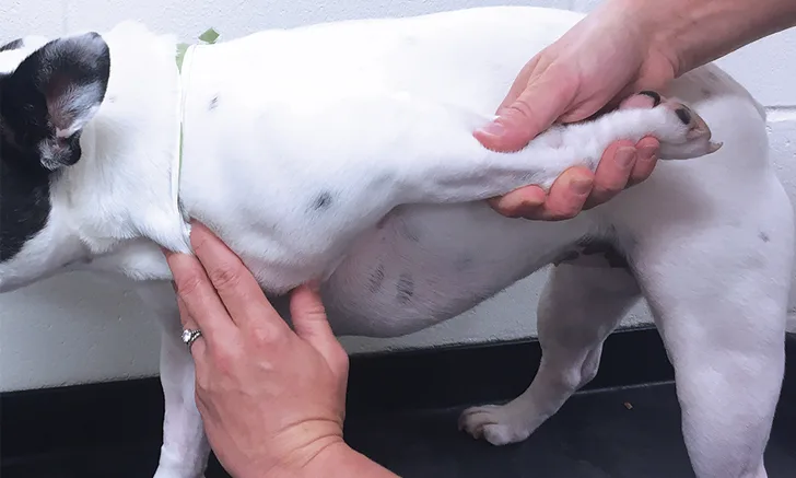
Biceps tenosynovitis is a common cause of forelimb lameness that most frequently affects medium- and large-breed dogs secondary to repetitive fatigue. It is characterized by inflammation of the biceps brachii tendon and the synovial sheath that envelops it in the shoulder joint (Figure 1).1,2 Clinical presentation often consists of a chronic progressive forelimb lameness that is worsened by exercise. The severity of lameness can vary from mild to nonweight-bearing, and atrophy of the supraspinatus and infraspinatus muscles is often present (Figure 2). Pain may be elicited during the biceps test, in which pressure is applied on the biceps tendon in the intertubercular groove when the shoulder is flexed and the elbow extended (Figure 3).1,2 Additional diagnostics (eg, ultrasonography, MRI, arthroscopy) are often needed to confirm diagnosis.1-3 Medical management (eg, rest, NSAIDs, physiotherapy) often results in resolution of mild lesions.1-3 Some dogs may require biceps tendon release or tenodesis to resolve pain and lameness.1-3

FIGURE 1
Arthroscopic image of the shoulder joint of a dog with biceps tenosynovitis. The biceps tendon (B) originates on the supraglenoid tuberosity of the scapula (A) and traverses distally through the bicipital groove of the humerus (closed arrowheads). Synovitis and increased vascularity of the tendon sheath are present (open arrowheads).
2. Achilles Tendon Strain
The Achilles tendon consists of 3 tendons: the superficial digital flexor tendon, the gastrocnemius tendon, and the combined tendon of the gracilis, semitendinosus, and biceps femoris muscles. Injury can occur secondary to acute trauma (ie, laceration, avulsion) or secondary to chronic degeneration, and clinical presentation depends on the severity of the injury and the tendons that have been injured.4,5 Mild strains may result only in lameness, pain, and swelling. If all 3 tendons have been severely compromised, the patient will walk with a complete plantigrade stance (Figure 4A); if only the gastrocnemius tendon has been injured, the patient will walk with a partial plantigrade stance (ie, increased flexion of the tarsus) with a noticeable flexion of the digits due to increased tension on the superficial digital flexor tendon (Figure 4B).4-6 Mild strains can be treated with medical management (eg, rest, NSAIDs, physiotherapy, orthotics); however, if a gait abnormality is present, surgical repair of the muscle–tendon unit is recommended.4-6


Two clinical presentations of Achilles tendon injury. A complete plantigrade stance with relaxed toe position, with complete rupture of the 3 components of the Achilles tendon (A), is present. Increased flexion angle of the tarsus with curling or flexion of the digits is present when the gastrocnemius tendon is ruptured and the superficial digital flexor tendon is intact (B).
3. Iliopsoas Muscle Strain
The iliopsoas muscle consists of the iliacus and psoas major muscle groups, which originate along the lumbar spine and ilium and insert on the lesser trochanter of the femur. Its main function is to flex and externally rotate the hip. Injury can occur secondary to an acute excessive force or repetitive use and/or trauma, resulting in a mild-to-severe pelvic limb lameness.7-10 Pain often can be elicited on examination by extension and internal rotation of the hip joint, abduction of the femur, and direct palpation of the muscle–tendon junction near the lesser trochanter.7-10 Because the femoral nerve runs through the iliopsoas muscle, some dogs that strain this muscle may also develop a peripheral neuropathy from compression of the nerve.8,9 Standard radiography can identify mineralization in the tendon, whereas ultrasonography, CT, and MRI are helpful for identifying early and subtle lesions and can help direct therapy (Figures 5 and 6).8-10 Mild-to-moderate acute lesions can often be treated with medical management (eg, rest, NSAIDs, physiotherapy, platelet-rich plasma injections).10 If the lesion is severe and results in fibrosis or contracture of the muscle, a partial tenectomy may be indicated.8,10

FIGURE 5
Ventrodorsal radiographic projection of a 9-year-old dog with right hindlimb lameness and hip pain. In addition to bilateral hip dysplasia and secondary osteoarthritis, mineralization is present within the right iliopsoas muscle near the tendon insertion on the lesser trochanter (arrowhead), which indicates a chronic strain injury.
4. Contracture of the Infraspinatus Muscle
Contracture of the infraspinatus muscle is most commonly seen in medium- and large-breed hunting dogs.11-14 Patients frequently have an initial forelimb lameness associated with an acute strain that resolves over several weeks. A characteristic forelimb gait abnormality then develops as the muscle irreversibly contracts.11-14 Because the infraspinatus muscle runs from the infraspinous fossa to the lateral aspect of the greater tubercle of the humerus, contracture results in the inability to extend the shoulder completely, and the limb is externally rotated and held in an abducted position.11-14 When walking, the limb is circumducted with the elbow partially flexed. This unique gait abnormality is easily observed when the patient walks up or down stairs (Figure 7). Surgical treatment is required and consists of a partial tenectomy of the infraspinatus muscle, which results in immediate improvement in gait abnormality and provides an excellent prognosis.11-14

Characteristic gait abnormality in a patient with infraspinatus muscle contracture. The limb is held in an abducted and externally rotated position and is circumducted with the elbow partially flexed when ambulating.
5. Luxation of the Superficial Digital Flexor Tendon
The superficial digital flexor tendon extends distal to the calcaneus, gliding over a bursa and the calcaneal tuber as it is supported by retinaculum on either side. Traumatic injury resulting in rupture of the retinaculum and medial or lateral displacement of the tendon has been reported in dogs and a cat.15,16 In Shetland sheepdogs, a hereditary basis has been established, and luxation is almost always lateral and caused by varying degrees of flattening of the lateral aspect of the calcaneal tuber.17 Depending on the inciting cause, clinical signs can include acute or chronic intermittent pelvic limb lameness and swelling around the calcaneal tuber.15-17 In some cases, the tendon can be luxated and reduced manually, with the tarsus held in extension.15,16 Medical management is ineffective for this condition.15-17 Prognosis is good to excellent following surgical treatment directed at repairing the torn retinaculum, imbrication of redundant retinaculum, and, in some cases, deepening the groove between the medial and lateral processes of the calcaneal tuber.15-17