Top 5 Indications for Fluid Therapy
Lisa L. Powell, DVM, DACVECC, BluePearl Veterinary Partners, Eden Prairie, Minnesota
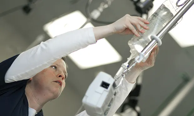
Fluid therapy, a mainstay treatment, is indicated in a variety of disease states that may result in mild dehydration to severe hypovolemic shock. Depending on the disease and its severity, fluids administered either subcutaneously or intravenously may be warranted. Fluids include crystalloids, synthetic colloids, and biological fluids (eg, red blood cells, plasma, concentrated albumin transfusions). This article focuses on crystalloid fluid therapy.
1. Dehydration
Dehydration, or the loss of fluid from the interstitial space, is most commonly caused by decreased intake and/or increased fluid loss from vomiting, diarrhea, or polyuria. The level of dehydration is often measured as a percentage of body weight via history and examination findings. Signs include decreased skin tenting, sunken eyes, depressed mentation, and tacky/dry mucous membranes. An estimate of dehydration is often not possible (or advisable) with just one variable; for example, false negatives may occur in very young patients, which have a higher percentage of total body water and tissue that is more elastic, and in obese patients. Signs often do not appear until dehydration is at least 5% of body weight; if a patient has a history of fluid loss and decreased intake but does not seem dehydrated, a 5% dehydration status should be applied.
To calculate daily maintenance fluid rates, the following equations can be used1:
Cats: 80 x body weight [kg]<sup0.75sup> or 2–3 mL/kg/hr
Dogs: 132 x body weight [kg]<sup0.75sup> or 2–6 mL/kg/hr
The deficit should be replaced over 8 to 24 hours, indicated by the patient’s clinical status, comorbidities (eg, heart murmur), and inpatient or outpatient status. For an inpatient, the deficit amount should be added to a base maintenance rate of isotonic crystalloid fluids and the patient monitored for ongoing losses that may require fluid rate adjustment. For an outpatient, the deficit may be administered subcutaneously and reassessed in 24 hours or as needed.
Calculating Crystalloid Fluid Rate in a Dehydrated Dog*
The following example shows how to calculate the maintenance rate and dehydration deficit of a 10-kg dog presenting with vomiting and diarrhea and with normal heart, lung, and kidney function:
Dehydration deficit
8% dehydration
body weight (10 kg) × % dehydration (0.08) x 1000 = 800 mL
Maintenance rate
132 × 10 kg0.75 = 742 mL/24 hours = 31 mL/hr
Replace dehydration deficit over 12 hours = 800/12 = 67 mL. Add this to the maintenance rate of 31 mL/hr = 67 + 31 = 98 mL/hr for the first 12 hours, then decrease to maintenance rate = 31 mL/hr.
*Refer to Fluid Therapy for the Emergent Small Animal Patient: Crystalloids, Colloids, and Albumin Products for available crystalloid fluid formations.2
2. Hypovolemia
Hypovolemia, or fluid loss from the intravascular space, may be caused by hemorrhage or extreme fluid loss from vomiting, diarrhea, polyuria, or prolonged decreased water intake. Signs include changes in perfusion indices: pale pink mucous membranes, prolonged capillary refill time (>3 seconds), tachycardia, decreased femoral pulse quality, depressed mentation. Patients are often hypotensive (ie, systolic blood pressure <90 mm Hg) and may have a metabolic acidosis and hyperlactatemia from poor perfusion resulting in lactic acidosis.
When an intravascular fluid deficit occurs, tissue perfusion decreases, resulting in tissue hypoxia and a change from aerobic to anaerobic metabolism. Lactate, a product of anaerobic metabolism, can be measured, which helps the veterinarian understand if the patient is responding to fluid resuscitation. Patients presenting in hypovolemic shock should be treated with IV isotonic crystalloid fluids via a bolus dose calculated on the patient’s shock volume (ie, 90 mL/kg [dogs]; 50–60 mL/kg [cats]). The general shock volume is often given as a fraction (ie, one-fourth to one-third of the total shock volume, or 30 mL/kg [dogs] and 10–20 mL/kg [cats]) over 15 to 30 minutes. After the bolus, the patient should be reassessed for therapy response, including heart rate, pulse quality, mentation, and lactate.
If perfusion abnormalities persist, an additional bolus may be used. Continued fluid therapy is usually warranted, depending on the underlying cause. Blood product transfusions may be indicated (10–20 mL/kg of packed RBCs, fresh frozen plasma, or whole blood, over 2–4 hours), depending on cardiovascular stability.
3. Distributive Shock
Distributive shock is caused by extreme vasodilation, including sepsis, systemic inflammatory response syndrome (SIRS), anaphylaxis, Addisonian crisis, and severe transfusion reactions and results in decreased tissue perfusion and tissue hypoxia. Treatment typically includes shock doses of IV fluid therapy. Inflammatory mediators released into circulation may cause significant vasodilation, and isotonic crystalloid fluid therapy alone is often ineffective. Combinations of isotonic crystalloids, synthetic colloids, hypertonic solutions, and blood products may also be needed, as well as vasopressors if hypotension or signs of shock persist. Empiric antibiotic therapy pending culture results is indicated if sepsis is suspected.
4. Kidney Failure
Administration of isotonic crystalloid fluids is common for patients with kidney disease. Newly diagnosed patients should receive IV fluid therapy to correct dehydration, improve perfusion, and promote diuresis while being monitored for body weight changes and urine output. Patients in polyuric kidney failure often require more fluids than the daily maintenance rate.
If the patient becomes oliguric (ie, urinates less than 1–2 mL/kg per hour after adequate rehydration) or does not urinate, fluid therapy may be contraindicated if the patient is overhydrated/edematous; depending on the underlying cause, diuretic therapy is usually indicated. Signs of overhydration include pleural effusion, pulmonary edema, abdominal effusion, increased body weight from decreased urine output, and excessive skin elasticity (ie, jelly- like movement when skin is tented). Intermittent subcutaneous fluid therapy is often prescribed for patients with chronic kidney disease and can be administered as an outpatient or at home, depending on the severity and stability of the disease.
5. Certain Toxicities
Many toxins are excreted, either fully or partially, via the kidneys, so fluid therapy with other treatment modalities is used to hasten toxin elimination. In some cases, such as NSAID toxicity (dogs and cats) and lily ingestion (cats), IV fluid therapy is recommended for 48 to 72 hours to help perfuse the kidneys and prevent acute kidney injury. The rate of administration should be 2 to 2.5 times higher than a typical maintenance rate to assure sufficient diuresis, as long as the patient’s clinical status does not contraindicate a high fluid rate.3
Conclusion
In most small animal presentations, IV administration is ideal. Clinical status, including improvement of hydration status, normalization of hypotension, and improved perfusion parameters should be monitored frequently in patients receiving fluid therapy because administration rates may need to be adjusted.