Top 5 Consequences of Brachycephaly
Anne Quain, BScVet (Hons), BVSc (Hons), MVetStud, GradCertEdStud (HigherEd), MANZCVS (Animal Welfare), DECAWBM (AWSEL), PhD, University of Sydney, Camperdown, New South Wales, Australia

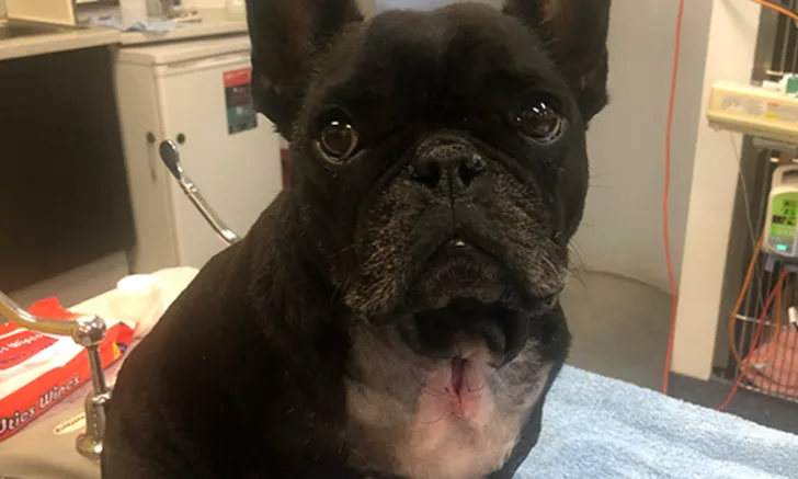
A French bulldog after removal of a temporary tracheostomy placed to bypass the upper airway due to obstruction triggered by severe heat stress. Some dogs require a permanent tracheostomy. Image courtesy of Dr. Ellie Leister, Veterinary Specialist Services
Brachycephaly (ie, shortening of the facial skeleton) is characteristic of some dog breeds (eg, French bulldogs, English bulldogs, pugs, Cavalier King Charles spaniels, Pekingese, Boston terriers). Brachycephalic conformation is associated with multiple health problems, some of which can be life-threatening and most of which are lifelong. Despite widespread publicity about these problems, popularity of these breeds as pets continues to increase.1 Awareness of the consequences of brachycephaly is important when advising pet owners about breed selection, advising breeders, and mitigating consequences for affected dogs. It should be noted that some syndromes are more common and/or severe in some brachycephalic breeds.
Following are the top 5 consequences of brachycephaly according to the author.
1. Brachycephalic Obstructive Airway Syndrome
Shortening of the facial skeleton leads to crowding and compression of the upper airway, resulting in upper airway resistance that increases the work of breathing and leads to air hunger during exertion; this syndrome is known as brachycephalic obstructive airway syndrome (BOAS).2 Anatomically, brachycephaly is associated with stenotic nares, hypertrophy of nasal turbinates, an elongated and thickened soft palate, a thickened tongue, everted laryngeal saccules, everted palatine tonsils, and a hypoplastic trachea; this can lead to partial or complete airway obstruction.3 Clinical signs range from mild stridor to severe dyspnea and collapse. Affected dogs may experience sleep apnea.4,5 Increased airway resistance can also result in soft tissue swelling and laryngeal collapse, exacerbating BOAS and potentially causing complete obstruction of the upper airway, necessitating emergency opening of the airway (Figure 1).6,7
Because owners are often unaware that BOAS indicates underlying pathology, and because upper airway noises may be considered normal for the breed, there can be delays in seeking veterinary care.8 Clinicians should advise owners on when to seek veterinary care and educate them on the sensitivity of brachycephalic breeds to heat stress (Figure 2).9
Early surgical correction is recommended to minimize progression of airway pathology in BOAS patients.10,11 Surgery typically addresses stenotic nares, elongated soft palate, everted laryngeal saccules, and palatine tonsils. Care must be taken when anesthetizing brachycephalic dogs with BOAS because of increased risk for complications (eg, regurgitation, aspiration pneumonia, respiratory distress).12,13 Perioperative administration of a prokinetic and a histamine blocker, minimal use of opioids, and recovery in an intensive care unit reduced the incidence of postoperative regurgitation in brachycephalic dogs.13

A bulldog with hyperthermia (body temperature, 107°F [41.5°C]) at an outdoor event. The dog was first cooled in tepid water then transported to a critical care facility. Brachycephalic dogs are prone to heat stress due to upper airway obstruction (ie, increasing inspiratory workload) and ineffective evaporative cooling.
2. Conditions Associated with Skeletal Abnormalities
Chiari-like malformation of the skull and craniocervical junction is most commonly seen in Cavalier King Charles spaniels, Brussels Griffons, and affenpinschers. Compression of neural tissue and disruption of CSF circulation can result and lead to development of fluid-filled cavities in the spinal cord (ie, syringomyelia [ie, neck scratcher’s disease]).14 Affected dogs may have concurrent ventriculomegaly,3 a painful condition most commonly seen in smaller brachycephalic breeds.14
Abnormalities of the vertebral column, including spondylosis deformans and vertebral malformations (eg, hemivertebrae), predispose brachycephalic dogs to intervertebral disk disease.15 Brachycephalic screw-tailed dogs (eg, French bulldogs, pugs, English bulldogs) are more commonly affected by spinal malformations, including kyphosis and scoliosis, which can increase the risk for intervertebral disk disease.3
Prognosis for affected dogs varies according to the severity of the underlying abnormality. Medical management with analgesic and anti-inflammatory drugs may relieve signs in mildly affected dogs. Surgery may be of benefit for some dogs; however, the prognosis is poor for dogs with marked scoliosis, intractable spinal pain, and/or neurologic signs refractory to medical management.3
3. Dental Disease
Brachycephaly is associated with dental malocclusion, overcrowding, and misalignment of teeth. A number of brachycephalic breeds have mandibular mesioclusion (ie, an undershot jaw), which is specified in breed standards; for example, French bulldog breed standards include an underjaw that is deep, square, broad, undershot, and well turned up.16
Malocclusion is associated with difficulty chewing food, temporomandibular joint dysfunction, trauma to soft tissue of the oral cavity, and premature tooth loss.17 Brachycephaly may also predispose dogs to dentigerous cysts, supernumerary incisors, and rotated, fused, or unerupted teeth (Figure 3).18 Some brachycephalic breeds (eg, boxers, bulldogs) may have prominent palatal rugae, in which plaque, hair, and food become trapped, leading to inflammation and development of granulomas; surgical correction may be required.18 Dental radiography is essential in the diagnosis and management of dentigerous cysts and supernumerary teeth. Owners should be advised to maintain dental hygiene and pursue regular dental examinations for their pet.

Oral cavity of a pug undergoing dental treatment. A rotated premolar and carnassial tooth, marked gingival recession and gingivitis, marked plaque and calculus, and fur entrapment can be seen.
4. GI Disease
Brachycephalic dogs are at increased risk for GI disease, including hiatal hernia, gastroesophageal reflux, esophagitis, delayed esophageal transit time, and redundant esophagus.19,20 French bulldogs have a higher incidence of hiatal hernia as compared with other dogs.20 Brachycephalic dogs presented with regurgitation and/or dysphagia are more likely to have esophageal motility disorders as compared with nonbrachycephalic dogs.21 Clinical signs of GI disease (eg, dysphagia, vomiting, regurgitation) are common in brachycephalic dogs with clinical upper respiratory tract disease.19 Of note, GI lesions were seen endoscopically in brachycephalic dogs that did not have GI signs.
Surgical management of respiratory disease may reduce GI signs.19,20 Because patients with hiatal hernia and esophageal disease are at greater risk for esophagitis and aspiration during and after anesthesia, patients with a history of regurgitation or reflux require close monitoring, and owners should be advised of increased risks.
5. Ocular & Ophthalmologic Disease
Brachycephalic breeds may have a variety of conformational defects (eg, medial canthal entropion, trichiasis, inappropriate tear fluid drainage leading to epiphora, qualitative and/or quantitative tear deficiencies, shallow orbits, proptosis, reduced corneal sensitivity) that compromise ocular health.22 These abnormalities increase the risk for ocular disease (including corneal ulcerations and erosions, vascular keratitis, pigmentary keratitis, corneal fibrosis, and keratoconjunctivitis sicca), leading to pain and vision deficits.22 Brachycephalic dogs are 11 to 20 times more likely to be affected by corneal ulcers than are nonbrachycephalic dogs.23,24 Nasal folds, visible sclera, and an increased eyelid aperture have been identified as risk factors.23
In some cases, surgical management (eg, medial canthoplasty) can reduce the risk for corneal exposure and irritation. Medical and surgical management may be required to manage acute and chronic conditions (eg, corneal ulceration). Careful attention should be paid to the ocular conformation of these dogs during examination so that owners can be advised accordingly.
Conclusion
Brachycephalic conformation predisposes dogs to respiratory, neurologic, dental, GI, ocular, and other disorders—including dermatologic abnormalities. Because these conditions affect the health and welfare of dogs, it is important they are not dismissed as normal for the breed.