Top 5 Canine Biliary Diseases
Stefanie M. DeMonaco, DVM, MS, DACVIM (SAIM), Virginia–Maryland College of Veterinary Medicine
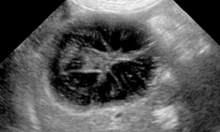
Ultrasonographic image of a GBM showing the classic kiwi-like appearance
The number of dogs diagnosed with biliary disease is increasing.1-5 Clinical signs and physical examination findings in dogs with biliary disease tend to be nonspecific and overlap with clinical signs of GI and systemic diseases; these can include anorexia, vomiting, abdominal pain, jaundice, and fever. Clinicopathologic abnormalities are similarly nonspecific and can include cholestatic to mixed liver enzyme elevations, hyperbilirubinemia, hypercholesterolemia, and neutrophilic leukocytosis.1,2,6-8 Diagnosis of biliary disease usually involves ultrasonography with or without collection of liver and bile samples. Treatment with urgent surgical care versus conservative medical management should be determined based on the cause and severity of disease.
Following are 5 of the most common canine biliary diseases according to the author.
1. Gallbladder Mucoceles
Gallbladder mucoceles (GBMs) result from an accumulation of semisolid mucus masses and inspissated bile in the gallbladder and are associated with high morbidity and mortality.3,6-11 Affected dogs are typically older (median age, 10 years) and of a predisposed breed (ie, cocker spaniel, Shetland sheepdog, miniature schnauzer, border terrier, Pomeranian).2,3,6-16 Additional risk factors for GBMs include gallbladder dysmotility, dyslipidemias, and endocrinopathies (eg, hyperadrenocorticism, hypothyroidism).14,17,18
Abdominal ultrasonography is key to diagnosis of GBMs. The classic description of GBMs is a kiwi-like appearance of intraluminal gallbladder contents with hyperechoic immobile striations of inspissated bile in hypoechoic mucus structures (Figure 1). Other ultrasonographic appearances of GBMs include echogenic immobile biliary sludge filling the gallbladder or a stellate pattern (Figure 2).6,10,11 These different GBM appearances on ultrasonographic images likely represent a continuum of early to mature mucoceles.6 Ultrasonography cannot be used alone to determine the clinical significance of GBMs or guide treatment decisions unless there is clear evidence of biliary rupture or obstruction that warrants urgent surgical intervention (see Gallbladder Rupture).1,3,10

Ultrasonographic image of a GBM displaying a stellate pattern
Preoperative biliary rupture and bile peritonitis can increase the risk for death, but some studies have shown that long-term survival (ie, 2-5 years) is possible in patients that survive the perioperative period.3,7,8,19,20 Common postoperative complications include pancreatitis and bile peritonitis.2,7,19 Patients with biliary infection at the time of rupture tend to have a higher mortality rate.
The best approach to treating GBMs (medical vs surgical) in dogs is controversial. When clinical signs and serum chemistry abnormalities (eg, increased ALP, γ-glutamyl transferase, ALT, and total bilirubin) are supportive of GBMs, cholecystectomy is generally recommended over medical management.1,6,10,21,22 A retrospective study found a longer survival time in dogs that underwent surgery as compared with those that received medical management.20 Medical therapy is best reserved for clinically inapparent cases of GBMs and when surgery is not an option. Medical therapy includes ursodiol, a low-fat diet, antibiotics, and treatment of concurrent diseases associated with GBMs (eg, hyperadrenocorticism, hypothyroidism, dyslipidemias) and, in most cases, is unlikely to resolve GBMs. A few cases of resolution or improvement with medical management have been reported, with other cases having static disease.2,10,23 Follow-up ultrasonography and serum chemistry profile performed within 2 to 3 months of diagnosis to assess response to treatment and identify complications are recommended, regardless of whether the patient is treated medically or surgically.
GALLBLADDER RUPTURE
Ultrasonography can help determine the presence of concurrent gallbladder rupture and/or extrahepatic biliary obstruction. Pericholecystic hyperechoic fat, pericholecystic fluid, a discontinuous gallbladder wall, and an unidentifiable discrete gallbladder with free-floating mucoceles in the peritoneum are supportive of gallbladder rupture.1,6,7 The specificity and sensitivity of ultrasonography in determining gallbladder rupture in dogs with GBMs varies from 91.7% to 100% and 56.1% to 85%, respectively.3,10
2. Extrahepatic Biliary Obstruction
The most common cause of extrahepatic biliary obstruction (EHBO) in dogs is pancreatitis. In acute pancreatitis patients, pancreatic edema and/or inflammation affecting the bile duct results in obstruction, whereas in chronic pancreatitis patients, fibrosis results in duct obstruction. Other causes can include GBMs, cholangiohepatitis, neoplasia, and cholelithiasis.4,13,24
Diagnosis of EHBO is usually made via ultrasonography and/or exploratory laparotomy. Ultrasonographic characteristics of EHBO include gallbladder enlargement, dilation of the cystic and/or bile ducts, and, in cases of obstruction lasting >5 days, intrahepatic duct dilation.9 Because ultrasonography may not always discern the cause of obstruction, surgery may be necessary to confirm biliary obstruction and further characterize and address the cause.9
Treatment of EHBO should be aimed toward addressing the underlying cause of obstruction and, if necessary, include biliary decompression. Whether surgical or ultrasound-guided percutaneous biliary decompression is necessary in patients with EHBO secondary to pancreatitis is controversial. Most dogs with EHBO secondary to pancreatitis improve with medical management as acute pancreatitis resolves. Serum chemistry abnormalities (eg, liver enzymes, bilirubin) can worsen despite improvement in clinical signs and should not be confused with worsening of the patient’s condition. Unpublished data suggest that bilirubin levels peak in dogs with pancreatitis-associated EHBO when clinical signs of pancreatitis are improving.
3. Cholecystitis
Cholecystitis can have acute or chronic presentations. Anorexia, vomiting, abdominal pain, and fever are typical signs of acute cholecystitis.25,26 Patients with chronic cholecystitis may have milder signs of chronic intermittent vomiting, anorexia, weight loss, and/or abdominal pain or no clinical signs at all. Cholecystitis may be present alone or in combination with cholangiohepatitis, which is typically characterized by chronic neutrophilic inflammation.4,5 Abdominal radiography can aid in the diagnosis of cholecystitis, particularly if emphysematous cholecystitis is present with a gas-filled gallbladder or gas opacities in the pericholecystic region. Nonspecific radiographic findings may reveal a right quadrant abdominal mass effect, poor serosal detail, and/or choleliths.9,27 The following abdominal ultrasonographic findings can be suggestive of cholecystitis and/or cholangiohepatitis: thickened, hyperechoic, irregular and/or laminar gallbladder wall; echogenic intraluminal contents; pericholecystic fluid or echogenic abdominal effusion; distended bile duct; and/or heterogeneous or hyperechoic hepatic parenchyma.4,9 Bile samples can be obtained with percutaneous ultrasound-guided cholecystocentesis to assess for inflammation, infectious agents, and culture and susceptibility. Culture and susceptibility testing is particularly important, as resistance can occur with empiric broad-spectrum antimicrobials.5 Common bacterial isolates include Escherichia coli, Enterococcus spp, Klebsiella spp, Clostridium spp, and Bacteroides spp.4,5,25,28
Treatment of cholecystitis includes medical management, but surgical intervention may be necessary depending on the severity of signs and gallbladder rupture. Cholecystectomy can reduce morbidity and mortality in dogs with cholangiohepatitis and/or cholecystitis.4 Medical therapy includes antimicrobial administration guided by either culture and susceptibility results or, when culture and susceptibility results are unavailable, empiric treatment against common isolates (eg, amoxicillin/clavulanic acid and enrofloxacin). Additional treatment options include ursodiol and supportive care.9,25 Cholecystectomy is typically the surgical treatment of choice when surgery is required and in cases in which only the gallbladder is affected.9,29
4. Cholelithiasis
Choleliths are stones in the biliary system and can have varying presentations (ie, mixed stones, pigment stones, cholesterol stones). In dogs with mixed stones, pigment stones are most commonly seen, with cholesterol stones being less frequent.9,30,31 Middle-aged to older, female, small-breed dogs are predisposed to choleliths, and an increased incidence of cholelithiasis has been observed in miniature poodles and miniature schnauzers.9,26,29,30,32,33 Choleliths are usually found incidentally on abdominal ultrasonographic images or necropsy and can lead to EHBO or cholecystitis.
Diagnosis is made via ultrasonography, which can detect stones >2 mm in size.9,26,27 Medical dissolution of choleliths is usually unsuccessful. Medical therapy includes ursodiol, S-adenosylmethionine, antimicrobials, vitamin E, and anti-inflammatory medications based on liver histopathology results (eg, chronic nonsuppurative hepatitis). Surgery is the treatment of choice in patients with concurrent cholecystitis and/or bile duct obstruction.
5. Biliary Neoplasia
Hepatobiliary neoplasia accounts for 0.6% to 1.3% of all canine neoplasms.34 Hepatocellular carcinoma is the most common form of hepatobiliary neoplasia, followed by biliary carcinoma. Labrador retrievers and female dogs are predisposed to biliary carcinomas.34-37 Ultrasonographic findings are nonspecific but can include a solitary mass or diffuse nodules with or without target lesions.38 Histopathology with or without immunohistochemical markers is necessary to confirm diagnosis.
The treatment of choice for biliary carcinomas is surgical resection unless the disease is diffuse or multifocal in nature. Overall survival is generally poor, with survival times typically being ≤6 months.34,39 Metastasis to regional lymph nodes and lungs occurs in ≤88% of dogs.35,36 Cholecystoduodenostomy can be performed in patients with secondary EHBO as a palliative option.