A 6-year-old spayed miniature poodle is presented for a 1-week history of lethargy, decreased appetite, and mild, bilateral epistaxis. She was previously healthy with no clinically significant abnormalities. The patient lives indoors and only goes outside to urinate and defecate. Monthly flea, tick, and heartworm preventives are current, and she does not receive any additional medications. Canine distemper virus, canine adenovirus, and rabies virus vaccines were administered 8 months prior to presentation. No interactions with other animals, snake bites, or insect stings are reported by the owner.
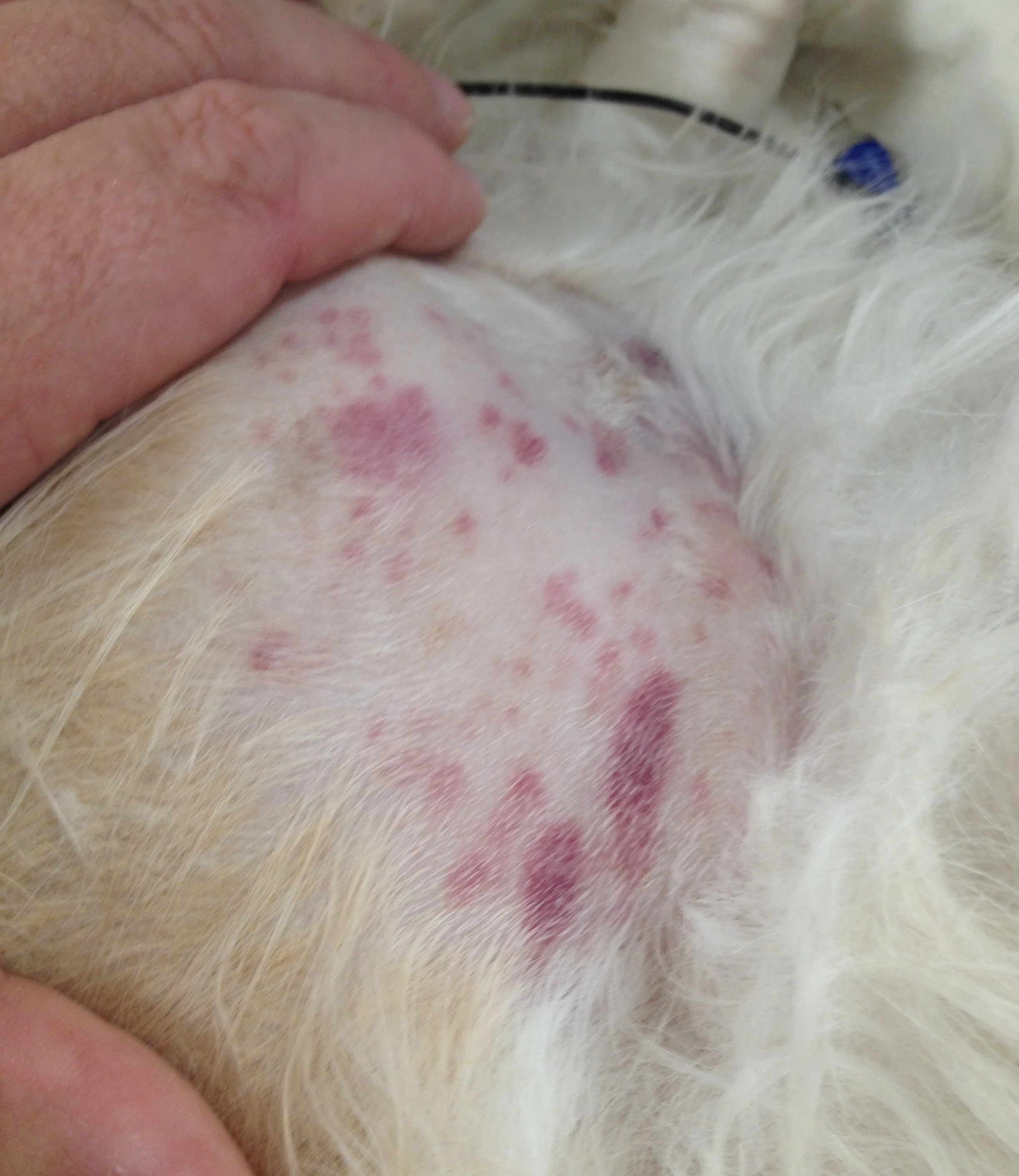
On physical examination, the patient is quiet, but alert and responsive. Mucous membranes are pink and moist. Temperature is 101.9°F (38.8°C), heart rate is 120 bpm, and respiratory rate is 24 breaths per minute. There is a mild amount of dried blood on both nares and moderate cutaneous petechiation and ecchymoses on the abdomen and ear pinnae (Figure).

FIGURE A Petechiation and ecchymoses on the abdomen
CBC results show a packed-cell volume of 36% (reference interval, 34%-60%), hematocrit of 37% (reference interval, 34%-60%), total leukocyte count of 21,500/µL (reference interval, 7,000-22,000/µL), and platelet count of 0 × 103/µL (reference interval, 160-650 × 103/µL). No platelet clumping is noted on blood smear evaluation, and the platelet estimate is 3 × 10³/µL (reference interval, 160-650 × 103/µL). Serum chemistry profile reveals no clinically significant abnormalities, and coagulation panel (prothrombin time and activated partial thromboplastin time) results are within normal reference intervals. A point-of-care ELISA test is negative for Dirofilaria immitis, Ehrlichia canis, E ewingii, Borrelia burgdorferi, Anaplasma phagocytophilum, and A platys. Results for additional infectious disease screening (titers and PCR analysis), including E canis, E ewingii, Rickettsia rickettsii, B burgdorferi, Babesia canis, and Bartonella henselae, are pending.
No significant abnormalities are detected on thoracic radiographs, abdominal radiographs, or abdominal ultrasound. Primary immune-mediated thrombocytopenia (ITP) is diagnosed based on patient history and clinical evidence.