Tail Mass in a Dog

A 4-year-old, male Labrador retriever presented with a persistent dermal nodule on the tail.

History and Physical Examination. The owner noticed a round alopecic lump on the dorsal surface and caudal aspect of the tail (which the dog licked frequently) a month earlier. The ulcerated, bloody, and crusty surface responded to a topical dermatologic cream but the nodule, which had a fluctuant-to-firm consistency, remained the same size.

ASK YOURSELF ...
What cytodiagnostic groups should be considered for this nodule as indicated by the history?
What microscopic findings are present in the smears from this patient?
What is the clinical significance of the cytologic findings in this dog?
Diagnosis: Deep pyoderma


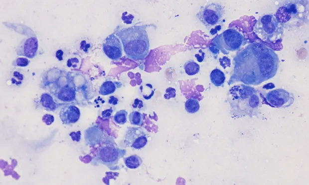
Figure 1. Note the basophilic epithelioid macrophages (black arrows) and binucleated macrophage (red arrow) in association with frequent neutrophils. Low numbers of lymphocytes are present; one is next to a fibroblast (black arrowhead)
Figure 2. A multinucleate macrophage (arrow) is surrounded by a mixture of neutrophils and epithelioid macrophages.
Figure 3. Nondegenerate (black arrows) and degenerate neutrophils (red arrows). Nuclear streaming cell debris (black arrowhead) and bacterial cocci (red arrowhead) are seen in the background. Inset shows neutrophils containing cocci.
Figure 4. Note the fragment of hair shaft or keratin in the center of the ball of neutrophjils (arrow) suggesting rupture of a hair follicle (furunculosis) (Wright's stain, 100x objective)
Cytologic EvaluationThe aspirate smear from the nodule has high cellularity, primarily inflammatory cells, and low numbers of anucleate squamous epithelium. Basophilic epithelioid macrophages (Figures 1 and 2) and few pale-staining phagocytic macrophages together account for more than 15% of the cell population. Occasional multinucleate macrophages (Figure 2) are noted. Neutrophils, both degenerate and nondegenerate, contain intracellular cocci bacteria (Figure 3). The background contains cellular debris and frequent clusters of three or more large cocci, some of which seem to adhere to the squamous epithelium.
Lymphocytes and fibroblasts are occasionally observed (Figure 1). A tight cluster of neutrophils is found arranged around a fragment hair shaft or keratin in the center (Figure 4). The cytologic interpretation is pyogranulomatous dermatitis with staphylococcal infection, presumed furunculosis.
Diagnostic ConsiderationsThe presence of monotypic bacteria suggests staphylococcal infection is this patient's primary disorder; however, it is prudent to rule out other conditions that can mimic the clinical presentation. Imprint cytologic evaluation of the skin surface is helpful to determine concurrent yeast infection with Malassezia species. Skin scrapings may reveal Demodex species infection. Histopathologic evaluation of the nodular lesion may be performed to further eliminate demodicosis, dermatophytosis, and follicular cyst rupture or adnexal tumor.
PyodermaThis common canine disease is defined as a pyogenic (or pus-producing) condition of the skin, most often associated with staphylococci (usually Staphylococcus intermedius). The organism may be found as a contaminant in normal canine mucous membranes, such as the nares and anus. Normal grooming may transfer the bacteria to the skin surface. Frequent licking of the skin encourages seeding into deeper portions, causing trauma to the hair shafts. Recently, two additional bacteria that show methicillin and fluoroquinolone resistance-Staphylococcus schleiferi subspecies schleiferi and coagulans-have been isolated from recurrent deep pyodermas. Culture with special biochemical testing may be warranted to determine the specific type of staphylococcosis.
Pyoderma is classified by the depth of bacterial involvement, which provides diagnostic and prognostic information. Surface pyoderma involves the epidermis without invasion into the stratum corneum or hair follicles and is associated with acute moist dermatitis ("hot spot" or pyotraumatic dermatitis) and skinfold pyoderma. Superficial pyoderma is a bacterial infection of the epidermis that forms pustules or inflammation on the outer or ostial portion of the hair follicle.
Deep pyoderma is more difficult to treat than the previous two types of pyoderma, as it extends into the dermis and hair follicles, destroying the hair follicle (furunculosis). The ruptured follicle wall releases fragments of the hair shaft as well as follicular keratins into the surrounding tissue, creating a foreign body reaction and pyogranulomatous inflammation in the dermis.
The hallmark of this inflammatory response is the presence of neutrophils, basophilic and pale-staining macrophages, and multinucleate macrophages (giant cells); this finding is suggestive of fungal and foreign body reactions. The young macrophages, which have moderate to deeply basophilic cytoplasm, lack evidence of active phagocytosis. The cytoplasmic borders of these macrophages often interdigitate with one another, creating an epithelial appearance, hence the name epithelioid macrophages. Young macrophages fuse with older macrophages to form multinucleate giant cells. When the nuclei of the multinucleate macrophage line up along the periphery of the cell, it is referred to as a Langhans giant cell and prompts consideration of an acid-fast bacterial infection (eg, Mycobacterium species). For current treatment recommendations, see Aids & Resources.
DID YOU ANSWER ...
• Cystic mass, inflammation, and neoplasia• Mixed inflammatory cell population and bacteria• The presence of neutrophils, macrophages, and multinucleate macrophages-referred to as pyogranulomatous inflammation-is suggestive of the type of fungal or foreign body reaction that may occur in a deep dermal lesion, such as deep folliculitis or furunculosis.