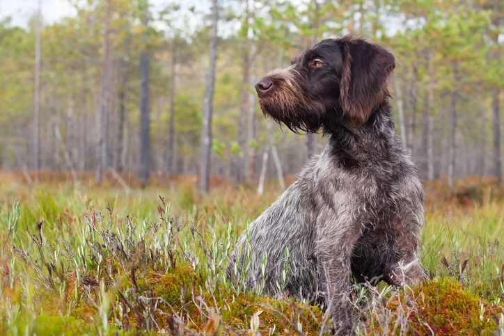
Jax, a 3-year-old intact male German wirehaired pointer, was presented for acute left thoracic limb lameness that developed after hunting pheasants ≈2 months prior. The owner did not observe a causative incident, and lameness did not improve with rest.
Physical Examination
On physical examination, Jax was bright, alert, and responsive. Vital parameters and thoracic auscultation were within normal limits. The left thoracic limb had moderate weight-bearing lameness, was mildly offloaded when the patient was standing, and had moderate muscle atrophy compared with the contralateral limb. Gross standing appearance of the limb revealed caudal swelling immediately proximal to the carpus (Figure 1). Palpation of the area revealed thickening on the caudal aspect of the antebrachium immediately proximal to the accessory carpal bone and elicited a mild pain response. Range of motion and stability in the mediolateral and craniocaudal planes of the left carpus were normal. No wounds or scars were visible over the region. The remainder of the physical examination was within normal limits.


Lateral (A) and caudocranial (B) photographs of the left antebrachium. Tendons on the caudal aspect of the distal antebrachium are thickened at the insertion of the flexor carpi ulnaris tendon on the accessory carpal bone (arrows). The mediolateral width of the normal right flexor carpi ulnaris tendon can be seen (star).
Diagnostics
Differential diagnoses for the soft tissue swelling, pain, and lameness included fracture, muscle or tendon strain, sprain, foreign body, and neoplasia.1 Although there was no known trauma, fracture was considered a possibility because field dogs commonly traverse uneven terrain and may step into holes, which can lead to significant supraphysiologic forces on the bones, ligaments, and tendons. In addition, sporting or working dogs can sustain fractures and soft tissue injuries secondary to repetitive strain injury to the bone.2-6
Lateral and craniocaudal radiographs of the left antebrachium did not reveal any fractures (Figure 2). A stressed, hyperextended lateral radiograph of the antebrachium to assess for palmar carpal stability was also normal.7


Lateral (A) and craniocaudal (B) radiographs of the left antebrachium. Thickening of the flexor carpi ulnaris tendon on the caudal aspect of the distal antebrachium is present (arrows). Hyperextended, stressed lateral radiograph of the carpus (C) does not show evidence of palmar ligament instability.
Based on the location of soft tissue swelling, a strain injury to the tendon of the flexor carpi ulnaris at the insertion on the accessory carpal bone was suspected. Musculoskeletal ultrasonography of the left and right antebrachium was performed for comparison and revealed significant tendon disruption of the insertions of the humeral and ulnar heads of the left flexor carpi ulnaris (Figure 3).8 Hyperechoic regions indicated fibrosis, which is typical of chronic injury.


Ultrasound images of the abnormal left flexor tendon (A) and contralateral normal right flexor tendon (B) at the insertion on the accessory carpal bone (arrows). The irregular fiber pattern and regions of hypoechoic and hyperechoic tendon in the left thoracic limb indicate tendon fiber disruption and fibrosis consistent with chronic injury. AC, accessory carpal bone
Diagnosis: Strain Injury of the Left Flexor Carpi Ulnaris Tendon
Treatment & Long-Term Management
Shockwave therapy and injections of platelet-rich plasma (PRP) in and around the abnormal flexor carpi ulnaris tendon were administered twice, 2 weeks apart. A closed-cell foam and synthetic rubber support
brace was fitted to the distal limb (from mid-radius to the metacarpal bones) for 4 weeks to provide support while allowing controlled loading of the tendon. Activity was limited to 5- to 10-minute walks on a leash 3 to 5 times per day, and rehabilitation was initiated with biweekly underwater treadmill sessions, class 4 laser therapy, acupuncture, and an exercise plan focused on isometric exercises and strengthening. An in-home therapy plan that included cross-frictional massage of the tendon (ie, firm massage applied perpendicular to the long axis of the tendon) 3 times daily, massage of the limb, and range-of-motion exercises for all joints in the limb was also implemented. Exercise restriction and physical rehabilitation therapy were advised until recheck examinations at 4 and 8 weeks.
Treatment at a Glance
Nonsurgical therapies (eg, laser therapy, shockwave therapy, PRP injections, acupuncture, massage) are appropriate if a tendon injury does not result in joint instability. Physical rehabilitation therapy should be implemented to maximize recovery.
Most tendon injuries can be treated with nonsurgical methods.
A brace can provide support during recovery to protect tendons and reduce risk for reinjury.
Prognosis & Outcome
At the 4-week recheck, lameness was improved, and thickening within the flexor carpi ulnaris tendon was decreased, but mild offloading of the left thoracic limb was still present.
At the 8-week recheck, Jax was fully weight-bearing on the left thoracic limb, and the carpus and elbow had full, pain-free range of motion. No pain response was elicited on palpation of the tendon, which was decreased in size but still ≈20% larger than the contralateral normal tendon. Ultrasonography revealed significantly improved tendon fiber orientation and organization. A gradual athletic physical reconditioning program was initiated, and Jax successfully resumed hunting pheasants with his owner 20 weeks after initial presentation.
Examination 1 year after the initial injury revealed mild thickening of the tendon but no pain or lameness.
Discussion
Tendons are essential structures in the musculoskeletal system because they connect muscles to bones and store and release energy from contracting muscles during locomotion, especially explosive movements.9 Tendon injuries are therefore common and should be a top differential for sporting or working dogs presented for lameness or performance issues. Early identification and treatment of these injuries is key to restoring tendon function and minimizing permanent damage (see Treatment at a Glance).9
Flexor Carpi Ulnaris Tendon
The flexor carpi ulnaris tendon is important for flexion and abduction of the carpus during ambulation and has antigravity functions during the stance phase of the gait.10 This musculotendinous unit has 2 muscular heads proximally, the humeral and ulnar heads, which function differently. The humeral head is stronger and likely has a greater role in locomotion because it has a slower contraction time and higher fatigue index compared with the ulnar head. Injury to one or both head-to-tendon insertions at the accessory carpal bone can lead to pain, lameness, and reduced athletic function. Sporting or working dogs undergo repeated stress to the flexor carpi ulnaris, which can lead to chronic tendon strain injuries.6 Lesions of the insertion of the flexor carpi ulnaris tendon have been suggested to result from low-grade strain followed by inadequate rest.3
Diagnosis of Tendon Injuries
Diagnosis of tendon injuries begins with standing and recumbent orthopedic examination. Orthogonal radiographs of the area of concern should be obtained, and stressed-view radiographs can facilitate objective assessment of joint instability. Musculoskeletal ultrasonography can be an effective point-of-care diagnostic tool to evaluate the location and severity of tendon injury. Comparison with the contralateral limb can help identify abnormal regions of tendon or bone. Additional diagnostic imaging modalities may include MRI to further evaluate soft tissues or CT to better evaluate bones for fractures or remodeling, which can be particularly helpful for the carpus or tarsus.11-13
Treatment of Tendon Injuries
Musculotendinous injuries typically do not require surgical repair unless they result in substantial loss of function.14-17 Basic principles for acute treatment of tendon injuries are cryotherapy (ie, ice), compression, rest, anti-inflammatory medications, and strengthening exercises.5 Chronic injuries may be treated with heat, rest, massage, and strengthening exercises.17 Other therapeutic modalities (eg, class 4 laser therapy, shockwave therapy, acupuncture, massage, orthobiologics [eg, PRP]) can improve the quality and speed of tendon healing through neovascularization and collagen synthesis and organization, and administration of growth factors can promote angiogenesis and cell proliferation, migration, and recruitment to heal damaged tissue.16,18-21 Shockwave therapy uses sound waves to stimulate neovascularization and increase collagen synthesis during early tendon healing,22 and PRP can introduce cytokines and growth factors that act individually and synergistically to promote healing within tendons.6 Support braces or orthotics can protect healing tendons from excessive forces while allowing controlled tendon and limb loading, which can improve the speed and extent of tendon healing and minimize limb atrophy.23-25 Braces and orthotics may be generic or customized to the patient, with some allowing a progressive and controlled dynamic range of joint motion via a hinge or adjustable straps to slowly increase the load on the injured tendon over time.
In cases of tendon function loss (eg, laceration or avulsion of the tendon from its insertion on bone), surgery to primarily repair or reattach the tendon may be indicated. Surgical repair is often protected for up to 8 weeks with a splint, cast, or orthotic to avoid gap formation that can occur during muscle contraction, followed by rehabilitation therapies and gradual increase in tendon loading.3,14,15
Prognosis for Tendon Injuries
Tendons heal slowly and never regain full strength due to scar tissue formation, which negatively impacts tendon structure and function. Following surgical repair, 56% of tendon strength returns after 6 weeks, and 79% returns after 1 year.26 Controlled tension across a healing tendon early in the course of healing orients fibers more optimally and results in greater biomechanical strength.23,24 A normal muscle applies the force of 25% to 33% of the tendon’s maximal strength during contraction; tendon healing may thus be acceptable for nonathletic function.27,28 In contrast, sporting or working dogs often use muscles and tendons at greater-than-normal loads and may thus be more likely to reinjure the tendon.
Conclusion
Identification and early treatment of tendon injuries are important to maximize treatment outcomes, minimize scar tissue formation, and allow return to function in sporting or working dogs. Surgical or nonsurgical treatments can be initiated depending on the severity of the injury, and prognosis can be very good if the injury is treated promptly, even though the tendon may not regain normal biomechanical strength. Physical rehabilitation therapy can improve patient comfort, maximize clinical outcomes, and potentially reduce the likelihood of reinjury.
Take-Home Messages
A high index of suspicion for tendon injuries is important when evaluating sporting or working dogs with lameness or performance issues.
Thorough history collection and orthopedic examination should be performed for sporting or working dogs presented with acute or chronic lameness. In addition to fractures, lacerations, and foreign bodies, tendon and ligament injuries should be considered because of the intensity of activities performed and uneven footing surfaces.
Radiography (including stressed-view projections) and musculoskeletal ultrasonography can be performed to evaluate bony structures and soft tissues. Ultrasonography can help assess tendon structure, integrity, severity of injury, and response to therapy. CT can further assess for concurrent osseous injuries. MRI can also be used if diagnosis is unclear; however, the expense is greater, and general anesthesia is required.
Tendon healing is slow, and reinjury is possible.