In the Literature
Forbes S, Bettenay S, Meertens NM, Wildermuth BE, Wildermuth K, Mueller RS. Diascopy and histopathological evaluation of nonblanching erythematous dermatoses in dogs. Vet Dermatol. 2024;35(3):255-262. doi:10.1111/vde.13230
The Research …
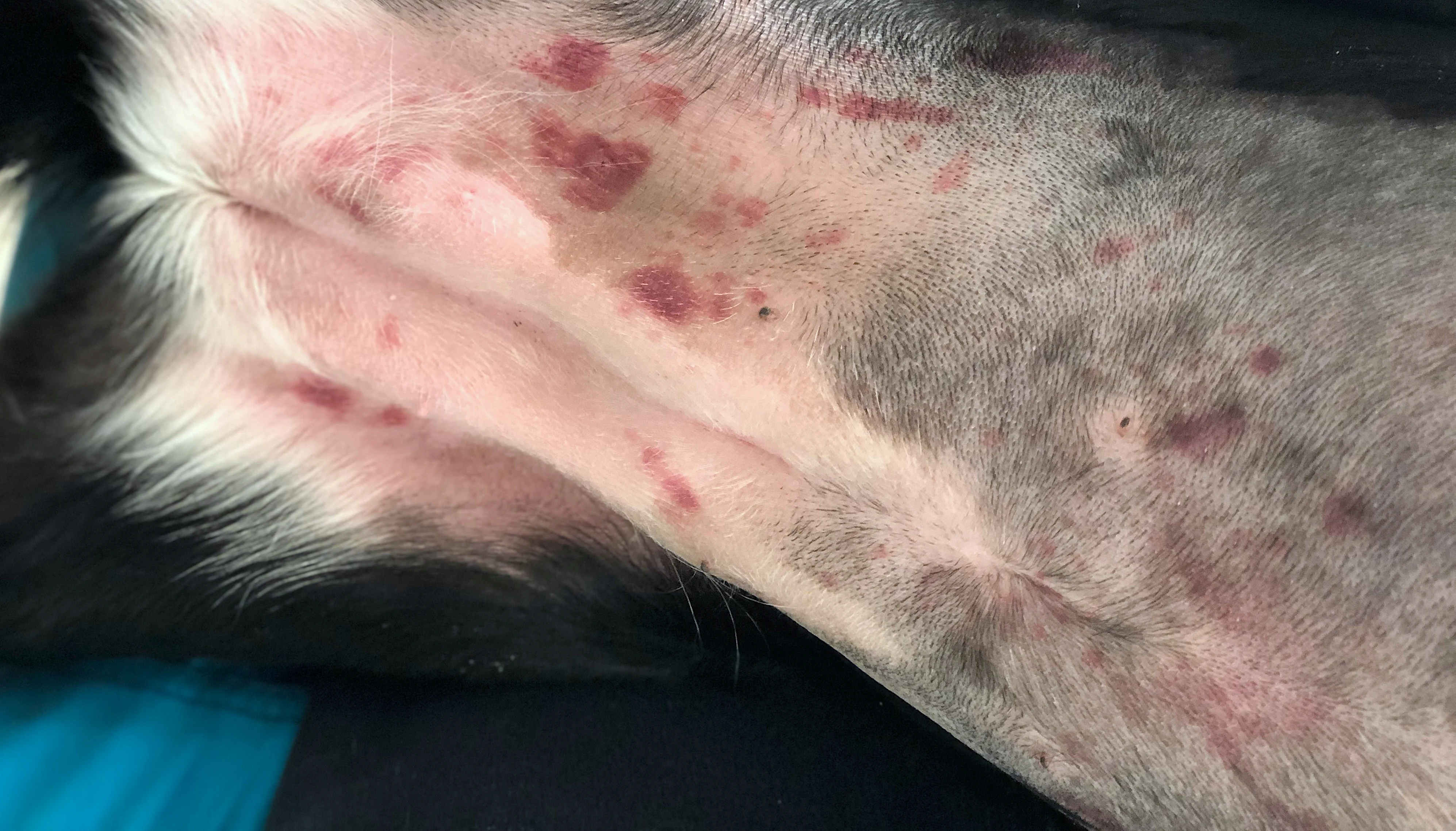
Erythema is a common clinical sign of skin disease. Various tools, including histopathology and diascopy, can help diagnose the underlying cause. Diascopy involves applying pressure to the skin, often with a microscope slide, to determine whether the skin blanches (ie, turns white; positive diascopy) or remains red (negative diascopy).1 In general, negative diascopy is associated with blood outside vessels (eg, hemorrhage, vascular damage), and positive diascopy is associated with blood inside dilated, intact vessels (eg, vasodilation due to inflammation).2 Results can help determine whether biopsy should be performed; conditions that cause vascular damage and hemorrhage are often definitively diagnosed via histopathology.
Negative diascopy is clinically described in patients with immune-mediated diseases (eg, immune-mediated vasculitis, cutaneous adverse drug eruptions, ischemic dermatopathy, erythema multiforme, canine acute eosinophilic dermatitis with edema [Well’s-like syndrome], canine granuloma pyogranuloma syndrome, canine acute neutrophilic dermatosis [Sweet’s-like syndrome]) and neoplastic conditions (eg, epitheliotropic cutaneous lymphoma); however, few published histopathologic reports verify hemorrhage and vascular changes in these patients.3-9
This retrospective study evaluated biopsies of dogs (n = 20) with nonblanching dermatoses to evaluate for vascular changes and hemorrhage. Histopathologic diagnoses identified 14 disease processes, with vasculopathy being most frequently diagnosed (6/20), followed by eosinophilic dermatitis (3/20) and cutaneous epitheliotropic T-cell lymphoma (2/20). Vascular changes, including perivascular edema, inflammatory cell infiltration of vessel walls, and swelling of endothelial cells, were identified in all dogs. Hemorrhage was also observed in 85% of cases. These results suggest nonblanching diascopy is associated with biopsy findings of hemorrhage and vascular damage.
… The Takeaways
Key pearls to put into practice:
Not all patients with negative diascopy had evidence of hemorrhage on histopathology. The authors speculated these cases (caused by calcinosis cutis, canine atopic dermatitis with insect bite reaction, and sebaceous adenitis with dermatophytosis) may have been false negatives that had some blanching on diascopy. Presence of false-negative cases may indicate that dividing diascopy results into blanching and nonblanching is an oversimplification of a spectrum of blanching and that diascopy is a subjective clinical test open to individual interpretation.
Some dogs (8/20) had diagnoses not previously associated with negative diascopy, including German shepherd dog pyoderma, mast cell tumor, hemangiosarcoma, exfoliative cutaneous lupus erythematosus, sebaceous adenitis, calcinosis cutis, and canine atopic dermatitis with insect bite reaction, suggesting many disease processes can lead to vasculopathy. Negative diascopy may thus not be as specific as initially perceived.
Results of this study support association between negative diascopy and conditions (eg, immune-mediated conditions, skin cancers) that typically require biopsy for diagnosis. Biopsy remains a reasonable recommendation in cases of negative diascopy.
You are reading 2-Minute Takeaways, a research summary resource presented by Clinician’s Brief. Clinician’s Brief does not conduct primary research.