Ringworm: Recognition & Client Education
Ringworm, or dermatophytosis, is a fungal infection of keratinized tissue with a characteristic ring-like appearance that affects both animals and humans. Three dermatophytes most commonly affect dogs and cats1:
Microsporum canis (transmitted through direct contact with an infected animal or human)
Microsporum gypseum (acquired through contact with infected soil)
Trichophyton mentagrophytes (typically transmitted through contact with rodents or their environment).
Related Article: Clinical Appearance & Diagnostics
Presentation
Although some ringworm infections may spontaneously resolve in healthy animals, management of the disease can often be frustrating and expensive.
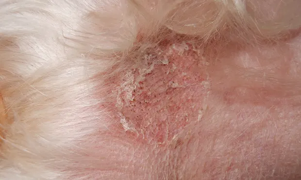
Ringworm’s classic presentation is a circular area of hair loss and crusted, slightly inflamed lesions that commonly appear on the face, ears, and legs. However, many ringworm cases do not fit this pattern and patients may present with a wide variety of signs (eg, generalized hair loss, nodules, hyperpigmentation [particularly seen in cats]).
Early diagnosis and treatment can lead to faster resolution of ringworm infection.
Diagnostics
Suspected lesions can be evaluated using a Wood’s lamp that has been allowed to warm for several minutes.
A yellow-green fluorescence will be seen on the hair shafts in areas positive for M canis; however, not all glowing areas (eg, crust, scales) indicate a positive test. The Wood’s lamp, therefore, should only be used to screen for ringworm because not all dermatophytes will fluoresce and false positives are common.
An in-house fungal culture using one of the following sample collection methods is the best procedure for diagnosing ringworm.<sup1-3 sup>
Remove hair and scale samples from the lesion edge using a clean hemostat; collect samples with positive fluorescence if the lesions were examined with a Wood’s lamp. Place the samples on a culture plate containing dermatophyte test media (DTM), lightly pressing the sample onto the media.
Brush the entire patient in the direction of the hair growth for 5 minutes with a new or sterile toothbrush and collect hairs and scale (ie, the MacKenzie toothbrush technique).2 This is most helpful for monitoring therapy response and/or cases where no discrete lesions are present. Gently transfer the sample to a DTM culture plate by lightly tapping the bristles onto the media.
Label the culture plates and incubate them lid-down at room temperature (25°C–30°C) away from direct light. Examine the culture plates daily, checking for growth or color change of the media. Dermatophyte colonies are unpigmented and white-to-buff-colored; media color change should occur before or in conjunction with fungal colony growth, although not every color change indicates dermatophytes.
If growth and color change are noted, examine the samples microscopically to identify the fungal organism3:
Place a drop of Methylene blue (or other dark) stain on a microscope slide.
Collect a sample from the culture plate by lightly touching the growth with the sticky side of a piece of clear tape.
Place the tape, sticky side down, on the microscope slide and examine at 10×–40×.
Examine for micro- and macroconidia to identify the dermatophyte.
Related Article: Talking to Clients About Zoonotic Diseases
Treatment
Treatment may include topical therapy, oral medication, or a combination. The prescribed treatments should be reviewed carefully with the client and provided in writing. It is important that clients understand how to properly follow the recommendations and are informed of any potential adverse effects, necessary laboratory monitoring, and the possibility of topical treatments staining the environment (most commonly with lime sulfur dips).
Treatment should be continued until at least one negative culture has been obtained and after the lesions have resolved.
Shaving may benefit long-haired patients. When clipping a patient, bathe with an antifungal shampoo or rinse, discard the hair, and immediately sanitize the clipper blades and treatment area to minimize exposure to other patients.
Treatment should be continued until at least one negative culture has been obtained and after the lesions have resolved. Clients must be reminded that dermatophytosis is zoonotic (ie, humans in contact with the patient are at risk of infection) and that they must not stop treatment until instructed by the veterinarian.
Environmental Control
Cleaning the patient’s environment is crucial to prevent recurrence of dermatophyte infections and to limit potential human exposure to the organism.4 Advise clients to:
Frequently wash (with hot water and detergent) any bedding, clothing, towels, and rugs the patient has contacted.
Dust all surfaces with disposable cloths.
Clean every area the patient has contacted, where possible, including floors, walls, and furniture, using a dilute household bleach solution (1:10 parts bleach:water); if possible, restrict the patient to one easy-to-clean area.
Sanitize or discard any combs, brushes, and toys.
Vacuum carpets frequently, disposing of the vacuum bag or emptying and cleaning the canister after each use.
Wash hands with soap and hot water and change clothing after contacting the patient.
Conclusion
Two considerations are key when managing patients with ringworm:
Treatment should begin as quickly as possible following the veterinarian’s diagnosis.
Clients must be educated about ringworm’s potential to spread to other household pets and to humans.
The zoonotic potential of this disease must not be underestimated, and frequent client communication and patient monitoring are the best ways to assure a successful outcome.
Editor’s note: Chris Keller worked in general veterinary practice until 2000 before transitioning to specialty practice. Animal Dermatology Center, where he serves as practice manager and veterinary technician, has specialized exclusively in dermatology since 2006.