Respiratory Distress in a Lovebird
Tiamo, a 5-year-old female lovebird, was presented for evaluation of labored breathing.
History. The bird had been breathing heavily for 5 days prior to presentation. During this time, droppings were darker than normal and the feet appeared reddish in color. In addition, 1 year ago her eggs began to appear misshapen.
Physical Examination. The bird was bright, alert, responsive, and mildly stressed. The feet were a dark shade of pink-red. The coelom was distended and felt doughy on palpation. She had mildly labored breathing on initial observation (92 breaths/min; normal, 50–60 breaths/min)1 with no respiratory sounds heard on auscultation. The remainder of the physical examination was unremarkable.
Laboratory Results. A complete blood count revealed mild hemoconcentration and leukocytosis (Table). Serum biochemical profile was within normal limits.

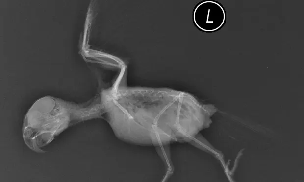
Radiographs. Plain whole-body radiographs (Figures 1 and 2) demonstrated diffuse loss of serosal detail in the coelomic cavity; the intestinal loops were gas-filled. The ventrodorsal view showed bilateral widening of the cardiohepatic silhouette. Caudal air sacs were obliterated by increased soft tissue opacity in the caudal coelom. Areas of bilaterally increased medullary opacity were seen in the humeri.
Ask Yourself...
What is the etiology of this patient’s clinical signs and diagnostic results?A. Lower respiratory tract diseaseB. Estrogen stimulation with probable reproductive diseaseC. Granulomatous mass in the caudal coelomD. Metabolic bone disease
Figure 1 (left): Ventrodorsal radiograph showing bilateral widening of the cardiohepatic silhouette.

Figure 2 (right): Lateral view showing bilaterally increased medullary opacity in the humeri.
Correct Answer:B. Estrogen stimulation with probable reproductive disease
Several findings in the history and examination of this patient suggest estrogen stimulation and reproductive tract disease. The eggs were misshapen, and there was an apparent caudal coelomic mass producing mild respiratory compromise. In addition, polyostotic hyperostosis was evident in the radiographs.
Discussion. Polyostotic hyperostosis (also called osteomyelosclerosis) is a nonspecific radiographic finding seen in female and occasionally male psittacine birds. It is most commonly reported in budgerigars and cockatiels, and cases have also been described in lovebirds and mynah birds.2-4
The condition is characterized by medullary opacity of several or many bones, obliterating the distinction between cortex and medulla on radiographs. The opacity results from new bone formation that is stimulated in part by estrogens from the maturing ovarian follicle.3
Hyperostosis likely represents a physiologic mechanism for egg production calcium storage because cyclic ossifications have been found to correlate with ovarian cycles in avians.3 Polyostotic hyperostosis is believed to result from excessive activation of this storage pathway, most often from hyperestrogenism.2-4 Laboratory correlation has been difficult to prove.
A study of 35 birds with hyperostosis did not find elevated levels of estrogens at euthanasia. Nevertheless, the majority of females had gonadal disorders, including ovarian cysts, oviductal adenomas and carcinomas, and ovarian granulosa cell tumors.3 Additional cases report similar gonadal cysts and neoplasms, including sertoli cell tumors in male budgerigars, suggesting a strong link with gonadal disorders.2-6
Diagnosis. Clinical findings in birds with hyperostosis are extremely variable, ranging from none to decreased activity and mild respiratory compromise in those with gonadal mass lesions. A soft tissue mass may be evident on physical examination. Ventral hernias, presumably due to hyperestrogenism, are also common.2,4 Radiographic assessment is critical to establish the diagnosis.
The differential diagnosis for increased medullary opacities of the long bones includes polyostotic hyperostosis, osteopetrosis, hypertrophic osteopathy, metabolic bone disease, and metastatic neoplasia.4-6 These other metabolic derangements of bone are often associated with periosteal new bone formation with thickened bones.2 The finding of a soft tissue mass in the coelomic cavity should prompt further evaluation for undeveloped eggs, egg yolk peritonitis, salpingitis, granulomatous disease, and cysts or neoplasms of the ovary or oviduct.4 Although not performed in this case, ultrasonography can be helpful in differentiating these conditions.
Treatment & Outcome. In our patient, the mass was presumed to be of gonadal origin, but financial constraints precluded further assessment. Large cysts can be surgically resected, or cyst contents can be aspirated. Salpingoopharectomy has been successfully performed for gonadal masses in birds; however, for many patients (including ours), surgery poses a significant risk of mortality.3,5-7
Medical management with leuprolide and human chorionic gonadotropin (HCG) has been used to manage hyperestrogenism.5,6 A decrease in ovarian stimulation by gonadotropins may help diminish the size of gonadal cysts and therefore reduce levels of estrogens. Our patient was treated with leuprolide (3 mg/kg) every 2 weeks. Although the radiographs remained unchanged at a 1-month follow-up, the bird had resolution of respiratory distress and improved activity.
Treatment at a Glance
• Hormonal control of hyperestrogenism: Leuprolide acetate, 3mg/kg IM Q 2 wk to inhibit estrogen secretion from the ovaries• Salpingoopharectomy will manage hypergonadism from cystic ovaries but can be a difficult procedure, especially in sick or small birds.
Take-Home Messages
• Not all problems that affect the respiratory tract originate there, particularly in birds. The differential diagnosis of respiratory distress must include primary respiratory disease as well as secondary effects from other conditions, including metabolic disease, anemia, and mass-producing lesions, including gonadal disorders. The unique anatomy of the avian respiratory tract, which contains multiple air sacs within the common coelomic cavity, makes it especially susceptible to mass lesions.• Full-body radiographs should always be performed for diagnostic purposes in the sick avian patient.• Documentation of increased medullary bone density in several or many bones, also known as polyostotic hyperostosis, should prompt evaluation for a source of excess estrogen stimulation, including gonadal tumors.• Hormonal therapy, including leuproloide and HCG, can reduce hyperestrogenism caused by underlying gonadal disease, often resulting in radiographic improvement of hyperostosis.