Respiratory Distress in a Cat
Salem, a 15-year-old, castrated male domestic shorthair was presented for evaluation of increased respiratory rate and effort.
History. Salem's medical history was unremarkable. He was one of two indoor cats that had been obtained as kittens from a neighbor. His vaccinations were up-to-date, and results of feline leukemia and FIV testing were negative. The respiratory distress had begun approximately 1 month before, with increased respiratory effort after exertion. Prior empiric therapy with enrofloxacin and prednisone transiently improved clinical signs; however, over the past 3 to 4 days, signs had become pronounced. In addition, Salem was anorexic and had been hiding.
Physical Examination. The cat was quiet, alert, and responsive. He was thin (body condition score 3 of 9). A grade 2/6 systolic murmur was present over the sternal border. Heart rate was 200 beats/min; pulse was moderately weak. Respiratory rate was 20 breaths/min, with moderately increased inspiratory effort. Increased bronchovesicular sounds were present, coupled with referred upper airway sounds. Abdominal palpation was unremarkable, except for slightly small kidneys. An enlarged thyroid gland was palpable on the left side of the cervical trachea.

Laboratory Work. A CBC, biochemical profile, urinalysis, thyroxine measurement, and chest radiographs (Figures 1 and 2) were performed. Results of the CBC and biochemical profile were normal, with the exception of high-normal levels of BUN (38 mg/dl; reference range, 6 to 35 mg/dl) and creatinine (2.1 mg/dl; reference range, 0.4 to 1.9 mg/dl). Urinalysis revealed diluted urine (USG 1.019) but was otherwise unremarkable. Serum thyroxine concentration was normal.
Differential Diagnosis. Problems identified include respiratory distress characterized by increased effort and referred upper airway sounds, heart murmur, and anorexia. Differential diagnoses for respiratory distress include upper airway obstruction (including mass or laryngeal paralysis), pulmonary thromboembolism, hyperthyroidism, or other metabolic or systemic disturbances. This case is challenging due to the lack of radiographic abnormalities consistent with most common causes of feline respiratory distress.


Diagnosis: Laryngeal squamous cell carcinoma
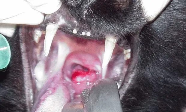
As a next step, because the problem was localized to the upper airway, an oral examination was performed under general anesthesia. Due to the concern of a laryngeal mass, supplies for an emergency tracheostomy were collected before sedation. Salem was preoxygenated for several minutes with flow-by oxygen and then anesthetized with propofol. Oral examination documented a mass (Figure 3). A temporary tracheostomy was done in the event of swelling and hemorrhage

and resultant airway obstruction after biopsy of the mass (Figure 4). Intubation could not be done using a standard endotracheal tube; therefore, a 5-French, red rubber catheter was inserted into the lumen of the airway to provide supplemental oxygen during placement of the temporary tracheostomy tube. A transoral biopsy was performed using biopsy forceps. In addition, because of concerns about neoplasia, partial arytenoidectomy was performed to try to reduce resistance to airflow.
Biopsy results revealed squamous cell carcinoma, a common laryngeal tumor. While distant metastasis is rare, the extent of the local invasion makes it difficult to treat.1 Salem underwent a permanent tracheostomy to bypass the upper airway. Permanent tracheostomies in cats are more likely to occlude than in dogs or other larger animals and require a committed owner. However, tracheostomies are able to alleviate respiratory distress associated with a laryngeal mass and are suitable as a palliative measure. Other treatment options for Salem included radiation therapy or chemotherapy. Piroxicam has been shown to have some antineoplastic properties against some carcinomas and is a potent analgesic, so it may also have a role in treatment of squamous cell carcinoma.2 Lymphosarcoma is also a common type of cancer associated with laryngeal masses. In cats, a nonneoplastic, granulomatous condition has also been appreciated.3
Salem lived for 2 months after the tracheostomy but was euthanized due to anorexia associated with tumor invasion in the caudal oropharynx. No difficulties were encountered with the tracheostomy site.
ASK YOURSELF . . .
Why was there an apparent response to prednisone and enrofloxacin?
What steps should be followed in pursing an oral examination?
DID YOU ANSWER ...
The prednisone may have decreased peritumor edema, resulting in initial improvement. Additionally, the enrofloxacin may have been effective if local infection from necrotic tissue was present.
The most important step in pursing an oral examination in a cat with a suspected upper airway obstruction is to be prepared for difficult intubation, including the potential need for emergency tracheostomy. An awake cat or dog may be able to breathe through a very small airway lumen; however, under anesthesia, this ability is inhibited. Intubation may be difficult and minutes lost locating supplies can cost the patient's life.
Upper airway obstruction due to neoplasia or laryngeal paralysis may cause respiratory distress. Neoplasia of the larynx usually results in narrowing of the laryngeal lumen and subsequent restriction of airflow. Laryngeal paralysis, although more common in dogs than in cats, has been described in cats.
Pulmonary thromboembolism can cause respiratory distress without radiographic abnormalities. It is very rare in cats, although it has been described.
Hyperthyroidism may result in tachypnea and restlessness due to a hypermetabolic state. Affected cats typically have extremely elevated thyroid levels. This patient's normal thyroid level excludes thyroid storm as a cause of tachypnea. Furthermore, inspiratory effort and referred sounds are inconsistent with thyroid storm.
Other metabolic or systemic disturbances. Metabolic acidosis occasionally accompanies severe renal failure, or diabetic ketoacidosis may result in compensatory tachypnea. Finally, intracranial disease, pain, or fever may also result in an altered breathing strategy.
RESPIRATORY DISTRESS IN A CAT • Elizabeth Rozanski
References
Feline oral neopl**asia: A ten-year survey. Stebbins KE, Morse CC, Goldschmidt MH. Vet Pathol 26:121-128, 1989.2. Evaluation of piroxicam for the treatment of oral squamous cell carcinoma in dogs.** Schmidt BR, Glickman NW, DeNicola DB, et al. JAVMA 218:1783-1786, 2001.
Obstructive inflammatory laryngeal disease in three cats. Tasker S, Foster DJ, Corcoran BM, et al. J Feline Med Surg 1:53-59, 1999.
Suggested Reading
Diseases of the larynx. Venker-van Haagen AJ. Vet Clin North Am Small Anim Pract 22:1155-1172, 1992.Laryngeal paralysis in cats: 16 cases (1990-1999). Schachter S, Norris CR. JAVMA 216:1100-1103, 2000.Tracheostomies in the management of canine and feline upper respiratory disease. Hedlund CS. Vet Clin North Am Small Anim Pract 24:873-886, 1994.