Quiz: Metastasis or No Metastasis: Radiography & Ultrasonography
Katherine Suter, DVM, BluePearl Specialty & Emergency Pet Hospital, Waltham, Massachusetts
David M. Schmidt, DVM, DACVR, Massachusetts Veterinary Referral Hospital, Woburn, Massachusetts
Clinicians should be able to determine the likelihood of metastatic disease based on the radiographic or sonographic appearance of lesions, as the presence of metastasis may affect the diagnostic plan, prognosis, and course of treatment. This quiz addresses the characteristics of metastatic lesions in the thoracic and abdominal cavities.
Conclusion
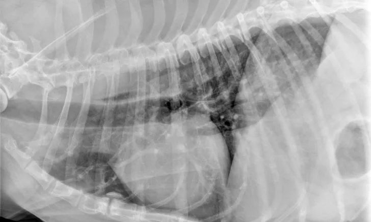
End-on pulmonary blood vessels are soft tissue opaque, perfectly round, superimposed over, and never larger than pulmonary blood vessels. Pulmonary osseous metaplasia (ie, osteomas) are mineral opaque, often irregular to angular, frequently small (1-3 mm), and not superimposed over vessels.
Metastatic pulmonary nodules are soft tissue opaque, usually round, variably sized (but >3-5 mm), and randomly distributed in the lungs. Nipples can mimic pulmonary nodules and are soft tissue opaque, often elongated (with only a distinctly margined caudal and ventral tip), and located ventrally.
Target lesions are round nodules with hypoechoic (dark) rims and hyperechoic (bright) centers on ultrasound and are highly suspicious for malignancy (>80%).2 Lymph nodes with round cell neoplasia are more likely to have hyperechoic perinodal fat.3 When appropriate, fine-needle aspiration or biopsy of a suspicious nodule should be performed to confirm diagnosis.