Peripheral pulse palpation can help determine circulatory and perfusion status and identify hypotension, arrhythmias, hyperdynamic states, peripheral vasoconstriction, and reduced cardiac output.1 Strong, palpable pulses synchronized to each heartbeat are normal and generally consistent with adequate cardiac output and intravascular volume. In patients with early compensated shock, however, pulse pressure and blood pressure can remain normal as other compensatory mechanisms (eg, tachycardia) develop. Once a reduction in pulse pressure is palpable, shock has progressed. Pulse pressure is the difference between systolic and diastolic arterial blood pressure and can be evaluated via digital palpation of pulse quality. Strength and quality of palpated pulses may allow evaluation of patient circulatory status and determination of underlying disease processes.
Detecting Pulses
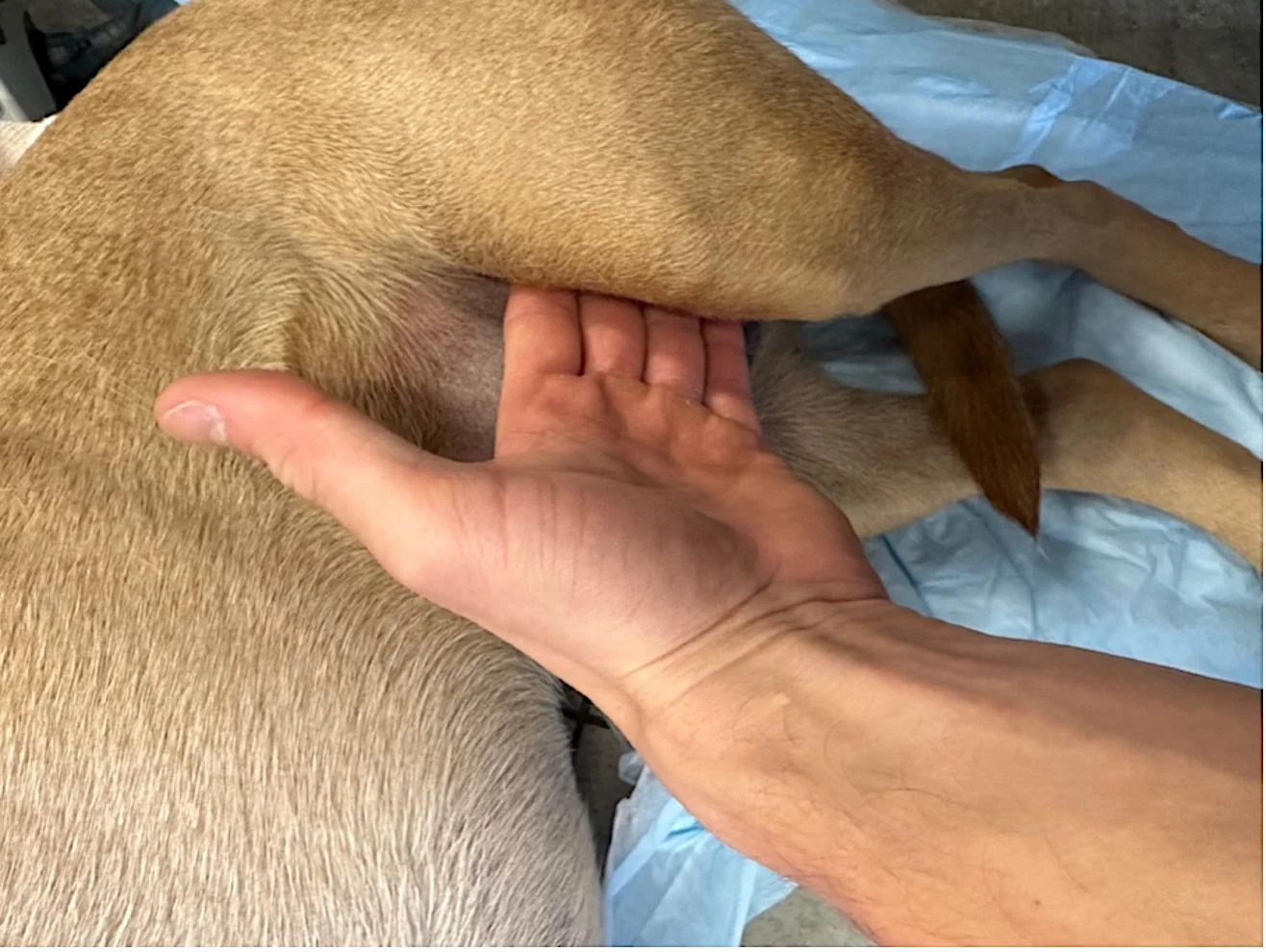
Femoral and dorsal pedal pulses should be palpated (ideally on both sides) while listening to the heart. The femoral artery is located in the femoral triangle (ie, area bordered by the inguinal ligament, medial border of the sartorius muscle, and medial border of the adductor longus muscle). This area should be compressed with the fingertips until no pulsations are felt, then gradually released until a pulse is palpated (Figure 1). The dorsal pedal artery is most easily palpated between the second and third metatarsal bones, just distal to the hock (Figure 2). Palpable dorsal metatarsal (pedal) pulses indicate a systolic pressure of ≥80 mm Hg.1,2 Absence of metatarsal pulses is highly suggestive of hypotension.3 Pulses that are irregular or asynchronous with each heartbeat can indicate an arrhythmia, and ECG monitoring should be performed to confirm and identify the arrhythmia and help determine optimal treatment.

Palpation of the femoral pulse in the inner thigh, approximately where the pelvic limb attaches to the body

FIGURE 2 Palpation of the dorsal pedal pulse, located on the dorsal proximal aspect of the metatarsal bones. The artery usually passes between metatarsals 2 and 3.
Pulse Rate & Rhythm
Pulse rate depends on patient species and status (eg, sleeping, stressed, excited, exercising). Consistently slow (ie, bradycardic) or fast (ie, tachycardic) pulses relative to normal should raise concern for cardiovascular abnormalities and warrant further investigation. Extracardiac causes can also affect heart rate but are not always indicative of shock; assessment of other perfusion parameters (eg, mucous membrane color, capillary refill time, mentation, distal extremity temperature) may therefore help determine whether shock is present.
Respiratory sinus arrhythmia manifests as an increased pulse rate during the inspiratory phase of respiration, followed by a slowed pulse rate during exhalation. This arrhythmia is not considered pathologic and is common in dogs, which typically have a high resting vagal tone, but uncommon in cats, which have a more sympathetically driven heart. Respiratory sinus arrythmia in a cat may indicate high vagal tone secondary to intracranial, respiratory, GI, or urogenital pathology.
A rapid and irregular pulse rate with variable intensity and pulse deficits is consistent with atrial fibrillation, brief bursts of supraventricular tachycardia, or ventricular tachycardia.4 Premature beats, characterized by a skipped pulse followed by a pulse stronger than others, may occur.
Pulse Character
Force and duration of arterial pulses on digital palpation may provide information about cardiovascular status and possible causes of a pulse abnormality.
Bounding/Hyperkinetic Pulses
Bounding/hyperkinetic pulses (ie, water hammer pulses) are generally snappy, have a rapid upstroke and descent sometimes described as tall and narrow, and are commonly associated with fever, exercise, aortic regurgitation, patent ductus arteriosus, severe hemodilutional anemia (eg, immune-mediated hemolytic anemia), or compensated vasodilatory shock. Bounding pulses are caused by increased heart rate and contractility, reduced blood volume, and stiffer arteries from compensatory vasoconstriction, producing a narrow pulse pressure with an amplitude (ie, pulse strength) higher than normal.5 Bounding femoral and metatarsal pulses should both be palpable.
Thready Pulses
Pulse pressure narrows, pulses become weaker, and pulse duration becomes shorter as hypovolemia progresses from mild to moderate and early decompensation occurs. Thready pulses may feel like a fine, mobile thread but can be difficult to palpate. Thready dorsal pedal pulses may or may not be palpable.
Weak Pulses
Weak pulses generally are palpable at the femoral artery (but not at the dorsal pedal artery due to low pulse amplitude), have a rapid rate, and may indicate decreased cardiac output from cardiogenic shock (eg, dilated cardiomyopathy, congestive heart failure, aortic stenosis, pericardial effusion) or an advanced stage of hypovolemic or distributive shock. Weak pulses are often a progression from thready pulses as the patient goes into decompensatory shock. Pulses may also feel weak as a result of local cellulitis or edema, which make palpation difficult.
Absent Pulses
Pulses may be absent due to circulatory collapse, decompensated shock, aortic embolism, or death. Absent pulses are not a sensitive measure of the return of spontaneous circulation in cardiopulmonary resuscitation and should not be relied on in cases of cardiac arrest.6
Conclusion
Pulse detection and characterization are valuable for monitoring intravascular volume status, cardiac status, and response to therapy. In general, pulse quality should improve as perfusion parameters return to normal. Changes in pulse character should prompt immediate blood pressure and ECG monitoring, especially if irregular pulses are present. A patient with perfusion deficits (eg, dull mentation, hypothermia, muddy/pale mucous membranes) can have both palpable dorsal pedal pulses and decompensated shock.3 Although pulse palpation can help evaluate cardiovascular status, findings should be considered in conjunction with patient clinical status to determine whether additional monitoring and diagnostics are warranted.