Proper Intraocular Pressure Measurement
Natalie Herring, LVT, VTS (Ophthalmology), Veterinary Specialty Center of Delaware, New Castle, Delaware

A thorough ophthalmic examination includes intraocular pressure (IOP) measurement, which aids in the diagnosis of glaucoma, uveitis, and other eye diseases. A veterinary nurse may perform this test or restrain the patient while the veterinarian performs the test.
Tonometry is used to measure IOP, and techniques for obtaining an accurate reading vary depending on the tonometer type. The indentation (Schiøtz), applanation (Tono-Pen VET), and rebound (Icare TonoVet) tonometers are the 3 most commonly used in veterinary medicine.
Related Articles2 Simple Tests for Assessing Ophthalmic HealthDry Eye in Dogs: When Good Glands Go BadKey Points in Complicated Canine Corneal Ulcers
An indentation tonometer uses a plunger and weight to indent the surface of the cornea. The indentation depth is measured against a scale and the value converted to a corresponding IOP. Indentation tonometry mostly has been replaced by applanation and rebound tonometry.
An applanation tonometer has a conical tip with a sensor that estimates IOP by measuring the force required to applanate (ie, flatten) an area of the cornea.
A rebound tonometer uses a magnetized metal probe that is launched at the cornea; the IOP is calculated by the induction current created when the probe rebounds.
Both applanation and rebound tonometers have similar disadvantages—the IOP measurement can be skewed by improper instrument use, improper placement on the cornea, and improper restraint. The rebound tonometer has some advantages compared with the applanation tonometer (eg, topical anesthetic is not required, the probe head is smaller, its settings are species-specific).
Video This video demonstrates how to perform an intraocular pressure examination using an applanation tonometer.
Following are step-by-step instructions for using applanation and rebound tonometers to measure IOP:
STEP 1 Setting Up the Tonometer
Always use a new, sterile tip cover or probe for each patient to avoid introducing contaminants in the patient’s eye.
Figure 1 Proper placement of an applanation tonometer tip cover. Photos courtesy of Veterinary Specialty Center of Delaware

To apply a new tip cover on an applanation tonometer, place the cover on the tip of the tonometer and gently roll it down to cover the entire tonometer tip. The cover should not be too tight or too loose because improper fit and placement can cause an inaccurate reading. (See Figure 1.) Calibrate an applanation tonometer at least daily, ideally between each patient, using the manual for calibration instructions.
Replace the metal probe of a rebound tonometer by placing a new probe directly into the tip slot with the footplate facing out from the unit. A magnet in the unit holds the probe in place. A rebound tonometer does not require calibration, but the correct patient species must be selected. The setting can be changed at any time.
Figure 2 Proper patient restraint. Photos courtesy of Veterinary Specialty Center of Delaware

STEP 2 Preparing the Patient
Place the patient in sternal recumbency, or in a comfortable sitting or standing position that encourages him or her to remain still. The team member restraining the patient should place one hand behind the patient’s head and the other hand around the lower jaw, taking care not to restrict the jugular veins with the hands or the patient’s collar. (See Figure 2.) Pressure on the jugular veins can increase IOP, particularly in brachycephalic breeds.1 Struggling with overly active or aggressive patients also may increase IOP. The team member taking the measurement must take care when manipulating the eyelids because excessive manipulation can cause a significant false increase in the measurement.2 When using an applanation tonometer, apply a topical anesthetic (eg, proparacaine hydrochloride 0.5%) to the patient’s eyes several minutes before performing the test. (See Figure 3.)
Topical anesthetic is not required when performing rebound tonometry but can be applied for patient comfort.

Figure 3 Proparacaine hydrochloride 0.5% ophthalmic solution. Photos courtesy of Veterinary Specialty Center of Delaware
STEP 3 Taking a Measurement
Hold the tonometer firmly when measuring IOP. To minimize the risk of dropping the unit, consider attaching a loop to the end of the tonometer that can be wrapped around the wrist.
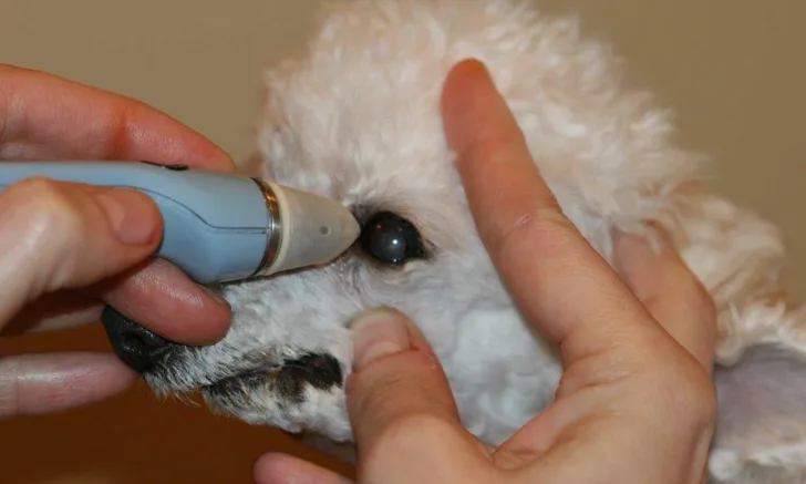
Apply pressure strictly against the cornea and avoid any pressure to the entire globe, as pressure on the globe may artifactually increase IOP.1 In brachycephalic breed dogs, globe location can make it difficult to apply pressure to the cornea only. Erroneous readings may be obtained when using either tonometer type if the tip or probe is positioned incorrectly during IOP measurement.

Figure 4 A properly placed Tono-Pen VET. Photos courtesy of Veterinary Specialty Center of Delaware
When using an applanation tonometer, hold the instrument at any angle, but be sure the tip is perpendicular to and within one-half inch of the patient’s cornea.3 (See Figure 4 & 5.) To obtain the measurement, ensure the footplate directly touches the cornea and then apply multiple light taps to the cornea. Approximately 3 repetitive taps are needed to take an average of the measurements. Applanation tonometers work by determining the force required to flatten a given area of the cornea.4
With a rebound tonometer, hold the device horizontally (ie, parallel to the floor) and position the tip of the probe perpendicular to the cornea, one-sixth to one-third of an inch from the cornea.5 This instrument uses a small probe suspended in the unit by a magnet. When taking a measurement, the unit must be held parallel to the floor because the probe will not properly fire in and out of the unit if the probe is held downward or upward. When the measurement button is pushed, the probe will be ejected from the unit and will gently rebound off the cornea. The handle position can be rotated without impacting readings so long as the probe direction remains horizontal and the distance to the cornea is consistent.4 Approximately 6 readings are required before this unit will provide an average.
Figure 5 A properly placed Icare TonoVet. Photos courtesy of Veterinary Specialty Center of Delaware

With each tonometer, the multiple readings are averaged to provide the IOP value. The normal IOP of most species ranges from 15 to 25 mmHg.1 Be suspicious about differences of more than a few mmHg between eyes.4
STEP 4 Cleaning and Storing
Clean the tonometer with a dry cloth to remove any debris. Always store the tonometer in its case with the tip cover or probe in place.
Periodically clean the tip of an applanation tonometer with compressed air according to the instruction manual.
Conclusion
IOP measurement is an integral component of the ophthalmic examination and aids in the diagnosis of eye disease. Applanation and rebound tonometry are most commonly used. Proper maintenance of instruments and use of correct techniques help ensure accurate readings are obtained.