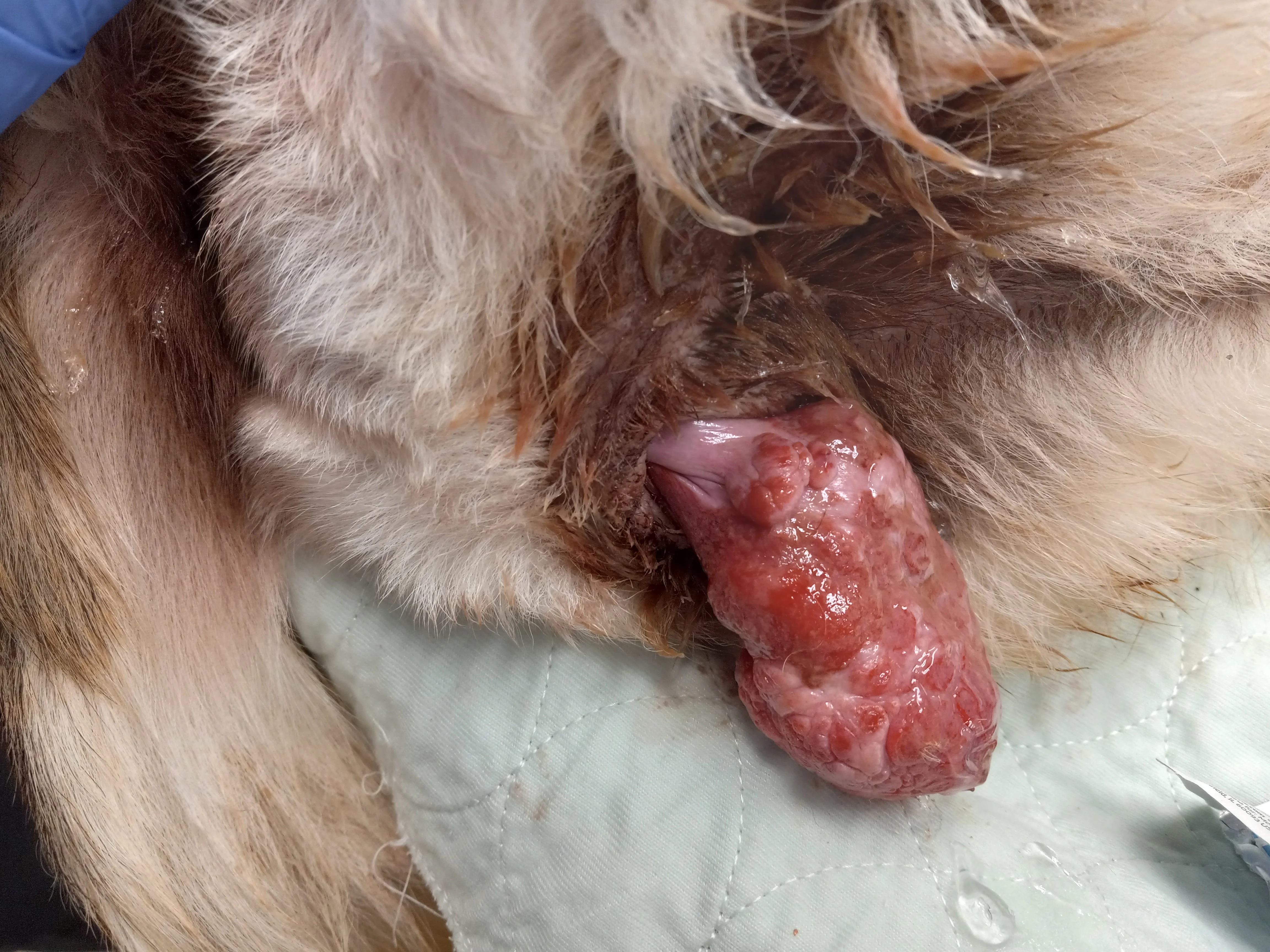
Determining Prolapse Type
Digital palpation can help determine whether a prolapse is uterine or vaginal. With a uterine prolapse, a finger can be passed completely around the tissue and reach the cervix. With a vaginal prolapse, the floor ± the circumference of the vagina prolapses; thus, a finger cannot be passed around the tissue on all sides, nor can the cervix be palpated in most situations.
Uterine prolapse occurs during or shortly after parturition and is likely related to relaxed tissues and contractions during labor. Vaginal prolapse occurs near parturition when progesterone levels are low and estrogen levels are high, after trauma, or secondary to a vaginal tumor. Vaginal prolapse can be confused with a vaginal fold prolapse (ie, edema of the vaginal mucosa that can worsen when estrogen levels are high during proestrus and estrus), a vaginal mass, or another type of tumor involving vaginal or urethral tissues.
Providing Emergency Treatment for a Prolapse
Prolapsed tissue should be cleaned and lubricated. To hold the tissue inside, loose sutures can be placed across the vaginal opening in an attempt to manually reduce the tissue and close the vaginal opening. Purse-string closures of the vagina can be traumatic to the tissue and should be avoided. Hyperosmotic solutions (eg, 50% dextrose, hypertonic saline) can be applied in an attempt to shrink prolapsed tissue. Cold water, sugar granules, and/or massage may help reduce tissue swelling and edema and make tissue easier to reduce. Overtly necrotic tissue should be debrided prior to manual reduction of tissue.
A complete uterine prolapse with all or most of the uterus protruding from the vagina may require emergent ovariohysterectomy to remove the prolapsed organ or laparotomy to place the organ back into the abdomen. Eventual (nonemergent) ovariohysterectomy to reduce estrogen levels is recommended to shrink existing prolapsed vaginal tissue and reduce risk for future vaginal prolapses.