Progressive Nonseasonal Pruritus in a Young Dog

A 2-year-old neutered male Parson Russell terrier was presented for a 1-year history of slowly progressive, nonseasonal pruritus.
History
The dog had received a tapering course of corticosteroids 2 months prior to presentation. This course began at 0.5 mg/kg every 24 hours and tapered over a period of 1 month with a good response. When asked to estimate the current severity of the dog’s pruritus, the owner gave a score of 9/10, reporting that the dog scratches 2 to 3 times per hour and seems to have difficulty sleeping because of intense itching. No other pets are in the household, and the dog is kept inside.
Physical Examination
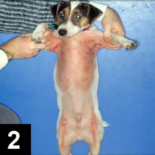
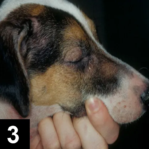
Moderate erythema, alopecia, and salivary staining were noted interdigitally on the palmar, plantar, and dorsal surfaces of all 4 feet. Mild erythema, occasional abrasions, and traumatic alopecia were present bilaterally in the axillae and over the ventral thorax, perineum, and medial aspect of the rear legs(Figures 1 and 2). Erythema was also noted over the muzzle, periocular region, and the concave surfaces of the pinnae and pinnal margins(Figure 3). No otic discharge was observed, and the ear canalsappeared normal. No ectoparasites were noted. The remainder of the physical examination was unremarkable.

Figure 1 (above): Erythema of the feet, axillae, periocular region, and muzzle

Figure 2 (above): Erythema and traumatic alopecia concentrated in axillae, ventral thorax, and medial stifles
Figu__re 3: Erythema of muzzle, pinnal margins, and periocular region
Ask Yourself...• When considering differential diagnoses, which parasitic, infectious, and allergic causes might explain this dog’s clinical signs?• Given the severity of the dog’s pruritus, what aspect of the patient history makes scabies an unlikely diagnosis?• Which diagnostic tests are most appropriate initially?
Did You Answer...• Malassezia dermatitis, dermatophytosis, superficial pyoderma, food allergy, atopy, and ectoparasites• Good response to corticosteroids• Skin scrapings and cytologic evaluation
Diagnosis: Atopy
Differential Diagnosis
Several differentials would exist in a case such as this. Typical to dermatology, the diagnosis is made by ruling out other diseases and by treating any secondary infections. Differential diagnoses would include Malassezia dermatitis, dermatophytosis, superficial pyoderma, food allergy, atopy, and ectoparasites.
Diagnostics
Skin scrapings showed no ectoparasites or fungal arthrospores. Impression smears of the axillae showed only epithelial cells and low numbers of degenerative neutrophils, but no Malassezia or bacterial organisms.
Given the patient’s age, the severity of the pruritus, and the results of the aforementioned diagnostics, the clinician suspected that food allergy or scabies was responsible for the patient’s condition. A commercial food elimination diet of strictly rabbit and potatoes was instituted in combination with an off-label trial of topical selamectin therapy (1 application every other week for 3 weeks). The latter trial was instituted despite the negative skin scraping because mites and fleas cannot be ruled out with 100% certainty without actually treating for them.
On reexamination 1 month later, no improvement was noted, ruling out the possibility of ectoparasites. The food elimination diet was continued strictly for another 6 weeks. When no improvement was noted after 10 weeks, the food elimination diet was discontinued and a diagnosis of atopy made.
Clinicians must remember that atopy is a diagnosis made only by exclusion because pruritus can be due to several different etiologies. For treatment to be effective for each disease process, it is important to make a final accurate diagnosis and not just treat the clinical signs.
Treatment
Treatment options for atopy are numerous. Generally, treatment for mild, seasonal cases of atopy can be initiated with antihistamines, essential fatty acids, and topical therapy. Topical therapy is important because the skin needs to be kept well hydrated, and newer treatments (such as topical products containing fatty acids) are aimed at replacing some of the ceramides that are considered to be inadequate in cases of atopy. In addition, bathing will help remove allergens that can be absorbed topically.
When these treatments do not adequately control clinical signs, other treatments should be considered.
Antiinflammatory dosages of corticosteroids are usually quite effective, at least initially, but have inherent risks associated with long-term or high-dose therapy. Another approach, immunotherapy with antigens selected based on allergy testing, can be beneficial and carries low risk for long-term side effects. Cyclosporine (Atopica, novartis.com) may also be considered.
It is important to discuss treatment thoroughly with the owner and select the option that best suits the dog with respect to age, size, and severity of clinical signs, as well as the owner’s economic and lifestyle limitations.
Outcome
The dog underwent intra-dermal testing to determine appropriate allergens for immunotherapy. During the initial month of immunotherapy, concurrent topical therapy was administered. Over the ensuing 4 months, the patient received two 2-week courses of prednisolone (0.25 m/kg Q 48 H) to maintain comfort. A 90% reduction in clinical signs was noted over the next year, and the remaining pruritus was managed with topical therapy and oral essential fatty acids.