Porcupine Quill Injury
A 7-year-old German shorthaired pointer was presented with acute respiratory distress and lethargy. Two weeks earlier, the dog had encountered a porcupine while hunting; quills were removed from the left forepaw, mouth, and sternal area. Ten days after the initial procedure, additional quills were removed from the thoracic region and left axilla. Following this procedure, the dog experienced an episode of retching, wheezing, and diarrhea. Lethargy and respiratory distress ensued 2 days later.
On presentation, the dog had bounding pulses, harsh lung sounds bilaterally, friction rubs in the right dorsal lung field, and an absence of lung sounds in the left ventral lung field. Thoracic ultrasonography findings appeared consistent with pericardial effusion. Radiographs revealed a pneumohydrothorax, pleural effusion, and possible pericardial effusion.
CT imaging revealed multiple quills in lungs, body wall, and mediastinal organs. Quills were surgically removed from the lungs, pulmonary artery, left ventricle, and mediastinum. Several partial- and full-lung lobectomies were required. The dog remained severely hypotensive postoperatively. An echocardiogram revealed presumptive porcupine quills remaining in the heart.
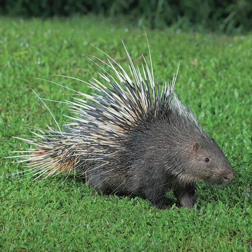
Porcupine quills can break off at the skin surface and, if not promptly removed, can migrate deep into body tissues.
Because of the need for cardiopulmonary bypass for further surgery and the grave prognosis, the dog was euthanized. Necropsy revealed a single porcupine quill in the left atrium above the mitral valve annulus. It extended across the aortic root and impinged on the coronary artery below the level of the aortic valve. Additional quills were discovered in the pectoral muscles, thoracic inlet, stomach, cecocolic junction, and in the main pulmonary artery of the right caudal lung lobe.
Commentary
Porcupine quill injuries are relatively common in certain regions. Quills are attached loosely to the porcupine’s body and are released on contact with an aggressor. The quills can break off at the skin surface and, if not promptly removed, can migrate deep into body tissues. If quill migration is suspected, CT is considered the gold standard for localizing foreign material, especially in the thoracic cavity.
In this case, echocardiography may have been useful to localize intracardiac quills preoperatively; however, both CT and echocardiography have limited sensitivity and specificity in discerning the exact location and extent of foreign material. This report emphasized the critical importance of complete quill removal immediately after the initial porcupine encounter to prevent complications associated with quill migration.—Sara A. Colopy, DVM, PhD, DACVS