Penile Mass in a Dog
A 2-year-old, castrated retriever crossbreed presented with a history of hemorrhagic discharge from the prepuce.
History
The dog was adopted from a shelter approximately 18 months before presentation; prior history was unknown. Approximately 3 months before presentation, the dog began to have hemorrhagic discharge from the prepuce. There was no evidence of hematuria, stranguria, or other urinary abnormalities. The patient’s appetite, activity level, and attitude remained normal, and the clients had not noticed any other abnormalities.
Examination
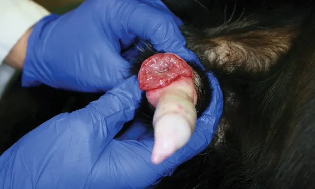
Figure 1: A lobulated, hemorrhagic, ulcerated mass of approximately 3 cm × 2 cm at the base of the penis. Image courtesy of Dr. Mary Lynn Higginbotham

The dog was bright and alert at presentation. Temperature, pulse, and respiratory rate were within normal limits. On palpation of the prepuce, a solitary mass was discovered at the base of the penis. Extension of the penis from the prepuce revealed a lobulated, ulcerated, friable, and hemorrhagic mass of approximately 3 cm × 2 cm (Figure 1).
There were no other clinically significant findings. An impression smear was submitted for cytological evaluation (Figure 2).

Figure 2: Impression smear of mass at the base of the penis. (Modified Wright’s stain, 200× original magnification [A]; Modified Wright’s stain, 600× original magnification [B])
Cytologic Description
The impression smears (Figures 2–5) were of high nucleated cellularity with scattered RBCs in the background. Most nucleated cells were round cells with rounded, eccentrically placed nuclei with a coarse chromatin pattern and prominent, sometimes multiple, nucleoli. Cytoplasms were basophilic and occasionally contained discrete small vacuoles. Anisocytosis and anisokaryosis were mild. Mitotic figures were scattered. Occasional neutrophils and eosinophils and rare macrophages were seen among neoplastic cells.
Figure 3: Most nucleated cells are neoplastic transmissible venereal tumor (TVT) round cells with round, eccentrically placed nuclei. Anisocytosis and anisokaryosis in this round cell population are mild. Mitotic figures (red arrows) were scattered throughout the image. Neutrophils (black arrows) and eosinophils (yellow arrows) are also evident. RBCs are scattered in the background. (Modified Wright’s stain, 200× original magnification)

Figure 4: The TVT cells have eccentrically placed nuclei that have a coarse chromatin pattern and prominent, sometimes multiple, nucleoli. Cytoplasms are basophilic and sometimes contained discrete small vacuoles. Mitotic figures (red arrows), neutrophils (black arrow), eosinophils (yellow arrows), and macrophages (green arrows) are evident. RBCs are scattered in the background. (Modified Wright’s stain, 600× original magnification)

Figure 5: Note the characteristic discrete, cytoplasmic vacuoles (pink arrowheads) and prominent nucleoli (black arrowheads) of the TVT cells. Mitotic figures (red arrows), eosinophils (yellow arrow), and macrophages (green arrow) are evident. (Modified Wright’s stain, 1000× original magnification)

Ask Yourself
Would you categorize the predominant population of cells as round, spindle, or epithelial?
What other cytologic features (eg, chromatin, cytoplasmic features) in this predominant population help specifically identify the lesion?
Which are the mitotic figures? What is their prognostic significance in this lesion?
Diagnosis
Transmissible Venereal Tumor (TVT)
TVT is a transplantable and contagious neoplasm of dogs that is transferred easily via direct contact of neoplasm to mucosa during mating or licking, which results in transplantation of tumor cells.1 The most common site for this tumor is external genitalia; however, lesions can be found on the face, nose, and nasal passages (ie, from sniffing and licking).2 TVTs are most commonly seen in patients from tropical areas with many free-roaming dogs<sup1–3sup>; scattered pockets of disease are seen in the United States. The author has recently diagnosed several cases of TVT in which none of the dogs had a known history of travel outside of Kansas, and all were adopted from Kansas shelters.
Classic TVT signs include genital, facial, or nasal masses (any size; may be friable, ulcerated) with no other physical abnormalities. If the mass on the genitals is large enough, it can cause hematuria, dysuria, or excessive licking.5 In patients with a competent immune system, the tumor is typically benign and often regresses spontaneously.2 Malignant TVTs have been reported; however, it is estimated that less than 10% of TVTs metastasize.5 Various sites of metastasis have been reported (eg, lymph nodes, subcutaneous tissue, skin, spleen, tonsils, liver, eyes, oral mucosa, pituitary gland, peritoneum, brain, bone marrow).6
Treatment & Prognosis
Although tumors may regress, treatment is highly recommended because of the contagious nature and potential complications of secondary infection, stranguria, or dysuria. Chemotherapy with vincristine is preferred for 2 weeks beyond resolution. A CBC is performed before every treatment to monitor neutrophil counts. If the total neutrophil count is less than 3000 cells/µL, the treatment is delayed.4 If the neutrophil count is lower than 1500 cells/µL and the patient is otherwise normal, an oral broad-spectrum antibiotic could be used to prevent secondary infection. Hospitalization, IV fluids, and antibiotics may be needed if the patient is neutropenic, febrile, or otherwise clinically abnormal.4 Treatment and hospitalization are dependent on the severity of illness.

The prognosis is good even without treatment as most TVTs will spontaneously regress within 6 months.1 Treatment usually results in a long-term control rate of 90%. However, in cases with distant metastasis, the prognosis is guarded.5
Clinical Outcome
The dog was evaluated and found to be a good candidate for chemotherapy. Vincristine was administered at 0.6 mg/m2 IV q7d for 7 weeks. There was no evidence of tumor regrowth at 6-month follow up.
Did You Answer?
Q: Would you categorize the predominant population of cells as round, spindle, or epithelial?
A: The predominant population is round cells.
Q: What other cytologic features (eg, chromatin, cytoplasmic features) in this predominant population help specifically identify the lesion?
A: The eccentrically placed, round nuclei with coarse chromatin patterns together with the punctate cytoplasmic vacuoles are features consistent with the cells of TVT.
Q: Which are the mitotic figures? What is their prognostic significance in this lesion?
A: The number of mitotic figures (Figures 3–5, red arrows, previous page) seen in TVTs does not have prognostic significance.
MARK MORTON, DVM, is a clinical pathology intern at Kansas State University, where he obtained his DVM. Dr. Morton is currently pursuing his master’s degree in Kansas State University’s veterinary biomedical science program. His interests focus on exotic veterinary medicine and animal behavior, as well as the cytologic and hematologic identification and characterization of disease in domestic species.
LISA POHLMAN, DVM, MS, DACVP, is director of clinical pathology and associate professor at Kansas State University. An active teacher of residents and students, Dr. Pohlman frequently speaks at national and international conferences to provide CE to practicing veterinarians. Her interests are in the areas of shelter medicine, veterinary forensics, and canine and feline lymphoma, as well as the cytologic and hematologic identification and characterization of disease in domestic species. Dr. Pohlman obtained her DVM from University of Guelph, spent 3 years in small animal practice, and completed her residency and master’s degree at Auburn University.