Pelvic Limb Lameness in a Cat
Kaitlin N. Bahlmann, DVM, Exclusively Cats Veterinary Hospital, Waterford, Michigan
Steven J. Bailey, DVM, DABVP (Feline), Exclusively Cats Veterinary Hospital, Waterford, Michigan
Zsa Zsa, an 18-month-old, 8-lb (3.6-kg) spayed domestic longhair cat, was presented for a 2-week history of left pelvic limb lameness and decreased activity levels. She was an indoor-only cat and was current on annual vaccinations, and her owners reported no known trauma.
Physical Examination
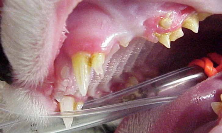
On physical examination, Zsa Zsa was bright, alert, and responsive. Vital parameters were within normal limits, except for an elevated heart rate (260 bpm). Oral examination revealed persistent deciduous teeth with coeruption of the permanent teeth, gingivitis, and calculus (Figure 1). Thoracic auscultation was unremarkable. Orthopedic examination revealed moderate left pelvic limb lameness. Pain and swelling were identified on palpation of the left stifle joint. Her BCS (5/9) and muscle condition score (3/3) were normal. The remainder of the clinical examination was unremarkable.


Persistent deciduous premolars in the right maxilla (A), left maxilla (B), right mandible (C), and left mandible (D). Coeruption of the deciduous and permanent maxillary canines, as well as marked gingivitis and calculus, particularly of the maxillary teeth, can be seen.
Diagnosis
Differential diagnoses for the patient’s stifle swelling, lameness, and pain included patellar luxation, atraumatic stress fracture of the patella, long bone fracture, soft-tissue or musculoskeletal injury (eg, cranial cruciate ligament injury), viral polyarthritis, and slipped capital femoral epiphysis. Inflammatory and neoplastic processes were considered less likely due to the patient’s age and absence of other clinical abnormalities. Patellar insufficiency fracture (ie, stress fracture) remained a likely differential due to the presence of persistent deciduous teeth and the possibility of osteogenic disease. Hypothyroidism was considered less likely due to the patient’s normal conformation and weight, as well as the absence of lethargy and anorexia.
The patient received buprenorphine (15 µg/kg IV) for analgesia, followed by an induction with alfaxalone (0.5 mg/kg IV) and general anesthesia with isoflurane to facilitate dental and orthopedic radiography. Dental radiographs revealed persistent deciduous teeth and impacted permanent teeth. Lateral and craniocaudal pelvic limb radiographs revealed a displaced left patellar fracture and a nondisplaced right patellar fracture (Figure 2).


Lateral radiographs of the left (A) and right (B) stifles show a complete, displaced left patellar fracture and a nondisplaced right patellar fracture, respectively.
Diagnosis: Feline Knees & Teeth Syndrome
Treatment & Management
Zsa Zsa was discharged on an NSAID (robenacoxib, 2 mg/kg PO for a total of 3 days) for pain management. Exercise restriction over the next several weeks was advised. She was returned to the clinic several days later for dental cleaning and nerve blocks, after which all persistent deciduous teeth and impacted permanent teeth were extracted. Buprenorphine (15 ug/kg IV) was administered, followed by an induction with alfaxalone (0.5 mg/kg IV). General anesthesia was maintained with isoflurane. Postoperative dental radiographs confirmed that no retained teeth or roots remained.
The patient was managed postoperatively with buprenorphine (15 µg/kg IV every 8-12 hours), and a transdermal fentanyl patch (12.5 µg/hour) was placed on the left lateral thorax. She began to eat within a few hours after recovery; left pelvic limb lameness persisted. Gabapentin (50 mg PO every 12 hours) was prescribed for multimodal analgesia.
TREATMENT AT A GLANCE
A thorough orthopedic examination should be performed on all cats with persistent deciduous teeth.
Careful extraction of retained deciduous teeth or impacted adult teeth is critical.
Staged dental extractions may be warranted to avoid iatrogenic mandibular fracture.
Conservative management with exercise restriction and pain management is generally advised.
Surgical management of patellar fractures is typically discouraged due to the high rate of surgical failure.
Fractures of the acetabulum, tibia, and other bones can occur; serial radiography should be pursued depending on the progression of clinical signs.
Some cats may heal and adapt to fractures, whereas others may continue to have an altered, plantigrade gait.
Prognosis & Outcome
At the 3-week postoperative recheck, Zsa Zsa demonstrated decreased left pelvic limb lameness and reduced associated stifle swelling. Oral examination revealed appropriate healing of the gingiva. Continued exercise restriction over the following 6 weeks, with gradual return to previous activity, was recommended. At the 3-month recheck, she was ambulating normally and exhibited no residual lameness or stifle swelling. Repeat pelvic limb radiographs revealed unchanged bilateral patellar fractures. No additional therapy was recommended for the patellar fractures due to concerns of surgical failure.
Serial radiographs of the pelvic limbs were taken periodically to monitor progression of the patellar fractures. Comparative orthopedic radiographs (Figure 3) 2 and 8 years after initial presentation revealed persistence of patellar fractures, with fragmentation, progressive sclerosis, and osteophytosis of the left patella.


Radiographs of the patellar fractures taken 2 years (A, left stifle; B, right stifle) and 8 years (C, left stifle; D, right stifle) after initial presentation. Progressive fragmentation and osteophytosis of the left patella and displacement of the right patellar fragments can be seen.
Discussion
Feline knees and teeth syndrome is an association of nontraumatic patellar fractures and persistent deciduous teeth. This pathologic syndrome may include persistence of deciduous teeth, unerupted permanent teeth, and insufficiency fractures of the patella.1,2 Other fractures, including in the long bones and pelvis, and spinal abnormalities have also been reported.3,4 Pathologic fractures of the tibia and fibula can occur up to 10 years after diagnosis.2
Feline knees and teeth syndrome was first identified in the United States, but affected cats have since been identified in South America and the United Kingdom.2,5 This syndrome was believed to be a manifestation of osteogenesis imperfecta, but another mechanism is currently supported,6,7 as patellar fractures showing radiographic evidence of sclerosis and generalized osteosclerosis have been seen in both humans and cats.8 In humans, osteomyelitis of the jaw associated with osteopetrosis and dental pathology has been reported.8-10 In addition, fractures of the patella and other bones have been associated with generalized osteopetrosis.8-10 Patellar sclerosis has similarly been observed in some cats with knees and teeth syndrome.2,4,11
Feline knees and teeth syndrome is typically recognized in young cats, with male cats more frequently affected than female cats.2,12 The mean age of onset of pelvic limb lameness or radiographic diagnosis is 28 months (range, 4 months-8 years).5 Physical examination findings typically reveal persistent deciduous teeth, pelvic limb lameness, and swelling of the distal quadriceps muscle; concurrent paronychia has also been reported.2,4 Intraoral and whole-body orthopedic radiography, with particular attention given to the pelvic limbs, should be performed for evaluation. In some cases, lameness and quadricep swelling precede visible radiographic fractures.2,4
Failure of deciduous tooth exfoliation in cats is rare and most often the result of an altered eruption path of the permanent tooth (see Take-Home Messages). Persistence of deciduous cheek teeth was reported in 40 of 60 cats with patellar fractures in one case series.2 Another series of 191 cats with various fractures reported that 48% had dental anomalies possibly related to knees and teeth syndrome.4 Extraction of both the persistent deciduous teeth and the impacted adult teeth is key, as proliferative osteomyelitis, dentigerous cysts, and jaw deformation may develop when these teeth are left in situ (see Treatment at a Glance).2,11,13,14 A careful, meticulous technique is required to avoid mandibular fracture due to the space occupancy of the impacted teeth in the mandible/mandibular canal.2
Although surgical reduction of patellar fractures may seem like a good option, surgical failure was reported in 86% of cats with feline knees and teeth syndrome.15 Therefore, nonsurgical, conservative approaches should be considered.15 Fractures in these patients may heal or the patient may adapt to the fractures and function normally even without surgical management; other cats may develop a plantigrade gait.2 A study of cats with knees and teeth syndrome that developed humeral fractures lacked sufficient data to determine the long-term prognosis for surgical repair, suggesting that many of these fractures may heal with medical management alone.3
Most cats that sustain pathologic fractures of the patellae related to knees and teeth syndrome adapt with good return to function. Cats with persistent deciduous cheek teeth should be closely monitored because development of pathologic insufficiency fractures of the patellae and other bones may be anticipated.
TAKE-HOME MESSAGES
Feline knees and teeth syndrome should be considered in cats that have spontaneous onset of pelvic limb lameness.
Cats with persistent deciduous cheek teeth should be closely monitored, as pathologic (especially patellar) fractures can occur.
Cats with juvenile dental anomalies should be further evaluated with dental radiography.
Extraction of any persistent deciduous teeth and unerupted adult teeth is recommended to prevent osteomyelitis, pain, and jaw deformation.
Acute lameness associated with nontraumatic patellar fracture often occurs at ≈2 years of age in cats with feline knees and teeth syndrome.
Pelvic limb lameness and distal quadricep swelling may precede patellar fractures.
Radiography of both pelvic limbs should be performed, even if lameness is only noted on one side.
Surgical reduction of patellar fractures is not recommended due to the high rate of failure.
Persistent lameness and an altered gait are possible long-term consequences of feline knees and teeth syndrome.
Fractures of bones other than the patellae may occur and should be monitored.