Panosteitis
Mary Sarah Bergh, DVM, MS, DACVS, DACVSMR, Midwest Veterinary Specialists, Milwaukee, Wisconsin
Panosteitis is a disease of the medullary bone that begins with adipocyte degeneration, intramembranous ossification, and bony remodeling that results in medullary fibrosis and periosteal/endosteal new bone formation; it should be considered a differential for any lameness in a young dog.
Clinical Signs
Panosteitis usually affects rapidly growing large- and giant-breed dogs, typically between 5 and 12 months of age, although it has been reported in dogs as old as 5 years of age.1,2 Some studies have found that German shepherd dogs and male dogs are overrepresented.1-3
Panosteitis causes an acute onset of lameness unaffected by rest or activity. Lethargy and inappetence can be seen for a few days at onset. More than 1 bone may be affected at a time. Clinical signs can commonly regress spontaneously in 1 limb and then occur in other limbs, causing a characteristic shifting leg lameness.
Diagnosis
A thorough physical and orthopedic examination is important, as other orthopedic diseases (eg, osteochondrosis, hypertrophic osteodystrophy) may be similar to signs of panosteitis and, in some cases, occur concurrently.
Pain on direct palpation of the diaphysis of long bones is characteristic of panosteitis. The ulna is most commonly affected, followed in frequency by the radius and humerus.1 Radiographs should be obtained to confirm diagnosis and rule out other pathologies.
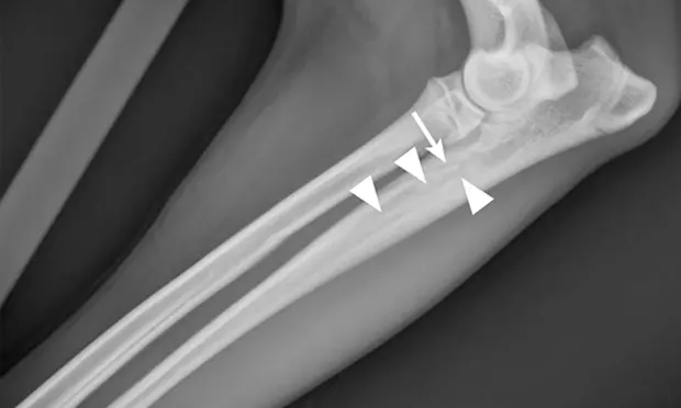
Radiographic signs of panosteitis frequently lag days to weeks behind clinical signs.1,3 The earliest radiographic sign of panosteitis is a decrease in opacity around the nutrient foramen. Later signs include an increase in mineral opacity within the medullary canal of long bones and loss of the normal trabecular bone pattern (Figure 1). Smooth periosteal and endosteal new bone may also be seen in more severe cases (Figure 2). Radiographs of the affected limb may be compared with those of the contralateral limb to assist in diagnosis. Nuclear scintigraphy may assist in diagnosis in cases in which radiographic changes have not yet developed.4
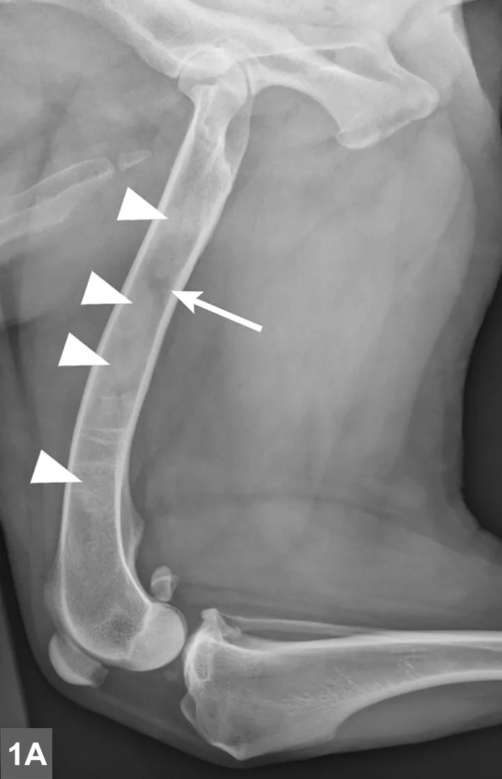
FIGURE 1A
Lateral radiographic projections of the femur (A) and ulna (B, next slide) in dogs with early signs of panosteitis. Note the radiolucency around the nutrient foramen (arrows) and increased opacity within the medullary canal (arrowheads) in both cases.
Treatment
Although the underlying pathogenesis of the condition is unknown, panosteitis is a self-limiting disease and resolves on its own. During episodes of pain and lameness, analgesia may be provided with NSAIDs, tramadol, narcotic analgesics, or gabapentin.
Prognosis
Episodes of pain and lameness from panosteitis last 2 to 5 weeks in each affected bone. It may recur until the patient is about 2 years of age, after which there are typically no long-term sequelae.1,3 For each recurrence of clinical signs, panosteitis should be confirmed through clinical examination and radiography to rule out other orthopedic causes.