Pain Management in a Dog Undergoing Orthopedic Surgery
Beatriz P. Monteiro, DVM, University of Montreal
Paulo V. Steagall, DVM, MS, PhD, DACVAA, Université de Montréal, Quebec, Canada

Fiona, an 8-month-old, 22-lb (10-kg) spayed Labrador retriever, was presented for severe lameness of the left pelvic limb and anorexia one day after being hit by a car.
Physical Examination
On examination, Fiona was depressed but responsive. Temperature was 100.4°F (38°C), heart rate was 130 bpm, and respiratory rate was 36 breaths/min. BCS was 5 of 9. Pain assessment with the short form of the Glasgow Composite Measure Pain Scale1 revealed a score of 8 of 24; scores greater than 6 of 24 indicate that interventional analgesia should be provided.1 Physical examination showed hematoma and marked edema in the region of the distal left femur. A fracture was noted on palpation. No other abnormalities were noted.
Diagnostics
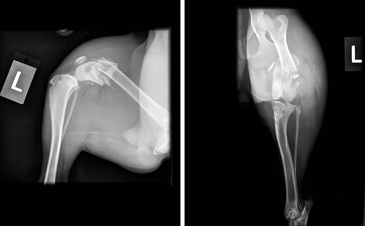
Hematocrit, total protein, and blood urea nitrogen values (obtained via dipstick) were 46%, 6 g/dL (60 g/L), and 5 to 15 mg/dL (1.78 to 5.36 mmol/L), respectively. Of note, if a patient remains hospitalized for more than 24 hours before fracture repair, hematocrit and total protein should be measured again on the day of surgery, as long bone fractures can be an important source of blood loss and consequent anemia. Abdominal-focused assessment with sonography for trauma (ie, aFAST) examination and thoracic radiographs disclosed no abnormalities. Pelvic radiographs showed a closed comminuted fracture of the distal left femur (Figure).

Mediolateral (left) and craniocaudal (right) radiographs of the left pelvic limb showing a comminuted fracture of the distal left femur. At least one transverse oblique fracture crosses the distal third of the femoral diaphysis. The distal fragment is displaced caudoproximally. A few bone fragments are visible near the fracture. The surrounding soft tissue is markedly enlarged in a circumferential pattern around the femur. No soft tissue gas, which would suggest an open fracture, is apparent.
Treatment & Management
The analgesic protocol was developed according to the WSAVA Global Pain Council guidelines.2 Hydromorphone (0.1 mg/kg IM) was administered for pain relief and moderate sedation, which allowed for additional diagnostic testing and IV catheter placement in preparation for surgical correction of the fracture. Anesthesia and analgesia were provided using a multimodal approach. Preventive analgesia was achieved as part of premedication through IV administration of a second dose of hydromorphone (0.05 mg/kg) in combination with dexmedetomidine (1 µg/kg).
Opioids provide optimal analgesia for acute pain and are safe when administered at clinical dosages3; they should typically be included in acute pain management protocols.3 Dexmedetomidine provides sedation, muscle relaxation, and some degree of analgesia; decreases volatile anesthetic requirements; and is part of a balanced anesthetic protocol. Opioids and α2-adrenergic receptor agonists produce synergistic effects that result in optimal analgesia.2
Anesthesia was induced using alfaxalone at 1 mg/kg IV to effect and maintained with isoflurane at 1.25% inspired concentration in 100% oxygen using a rebreathing system. Lumbosacral epidural anesthesia was provided with preservative-free morphine (0.1 mg/kg) and bupivacaine (1 mg/kg).
Local anesthetics provide excellent volatile anesthetic-sparing effects, blunt stress response to surgery, and reduce analgesic requirements in the perioperative period.4 Morphine produces long-lasting analgesic effects (16-24 hours) when administered via epidural route.3 Administration of local or regional anesthesia eliminates the need for continuous infusion of opioids, α2-adrenergic receptor agonists, and ketamine and/or lidocaine.2
Anesthesia and surgery proceeded uneventfully with no hypotensive events. The fracture was stabilized using internal fixation. Carprofen (4 mg/kg SC) was administered immediately before extubation. NSAIDs such as carprofen provide excellent perioperative analgesia and should be used for orthopedic procedures unless contraindicated.2,5 NSAIDs can be administered during the intraoperative period if patients are healthy, fluid therapy is administered, and blood pressure is monitored.5
While Fiona recovered from anesthesia, the surgical site was treated with cold therapy using ice packing for 15 to 20 minutes. Cold therapy is an inexpensive, nonpharmacologic therapy that can reduce swelling and provide analgesia for postoperative pain, particularly following orthopedic surgery.2
Fiona remained hospitalized for 48 hours. A soft, padded bandage was applied to the left pelvic limb, and she rested in a comfortable bed in a clean, quiet environment. For the first 36 hours after surgery, she received carprofen (4 mg/kg SC q24h) and hydromorphone (0.05 mg/kg IV q6-8h) for analgesia based on pain scores assessed hourly using the Glasgow Composite Measure Pain Scale. Food and water were available ad libitum. Fiona was taken on short walks using a sling support and received cold compression therapy (10 minutes 6 times a day) along with gentle massage of the affected limb (5 minutes 6 times a day).
Patient needs should be evaluated based on clinical status and welfare, including pain assessment, general comfort, and anxiety level. Optimal pain management can be provided using pharmacologic and nonpharmacologic options.2 Nonpharmacologic modalities (eg, cold and hot compression therapy, massage, a warm bed, frequent walks on sling support) are important aspects of pain management following orthopedic surgery. As the patient begins to heal, additional therapies (eg, range-of-motion exercises) can be added and the frequency of other modalities decreased.
Approximately 36 hours after surgery, Fiona was bright, alert, and friendly. Pain was not elicited on palpation of the surgical site. She was discharged on gabapentin (10 mg/kg PO q12h for 4 days) and carprofen (4 mg/kg PO q24h for 7 days). Instructions were provided for cold and hot compression therapy (ie, cold compressions for 10 minutes 2-3 times a day for 1 week followed by hot compressions for 10 minutes 2-3 times a day for 4 weeks) and range-of-motion exercises (ie, 10 repetitions of flexion and extension 2-3 times a day).
Treatment at a Glance
Preoperative: Hydromorphone (0.1 mg/kg IM); premedication with a combination of hydromorphone (0.05 mg/kg IV) and dexmedetomidine (1 µg/kg IV)
Induction & Maintenance: Alfaxalone (1 mg/kg IV to effect); isoflurane 1.25% inspired concentration in 100% oxygen using a rebreathing system
Intraoperative: Bupivacaine (1 mg/kg) and morphine (0.1 mg/kg) lumbosacral epidural; carprofen (4 mg/kg SC) administered immediately before extubation
Immediate Postoperative (First 36 Hours): Hydromorphone (0.05 mg/kg IV q6-8h depending on pain evaluation), carprofen (4 mg/kg SC q24h), cold compressions (10 minutes q4h), massage (5 minutes q4h), a warm bed, and frequent walks on a sling support
At Home: Gabapentin (10 mg/kg PO q12h for 4 days), carprofen (4 mg/kg PO q24h for 7 days), cold compressions (10 minutes q8-12h for 1 week) followed by hot compressions (10 minutes q8-12h for 4 weeks), and range-of-motion exercises (10 repetitions of flexion and extension q8-12h)
Prognosis & Outcome
Prognosis was good to excellent, provided the postoperative recommendations were strictly followed. At 8- and 16-week follow-up examinations, Fiona was recovering well. She was eating, drinking, urinating, and defecating normally. The owners were able to administer her medications and conduct the cold and hot compression therapy in the weeks following discharge.
Physical examination was unremarkable, and palpation of the fracture region elicited what appeared to be only mild discomfort. Moderate muscle atrophy was visible in the left thigh as compared with the right thigh. Radiographs of the left femur showed a well-aligned fracture that was healing appropriately. Instructions were given for a gradual return to physical activity over the following 6 to 8 weeks.
Take-Home Points
Fractures cause moderate-to-severe pain. Aggressive perioperative analgesia should be provided using a multimodal preventive approach.
The term preventive analgesia has replaced pre-emptive analgesia. Preventive analgesia encompasses all perioperative efforts to decrease postoperative pain and consumption of analgesic agents.
Multimodal analgesia involves use of different classes of analgesic agents to achieve synergistic effects and decrease adverse events.
Local and regional anesthetic techniques are cost-effective options for providing analgesia.
Nonpharmacologic approaches complement use of analgesic agents in pain management.