Otitis Externa
A 5-year-old, neutered male, mixed-breed dog was presented with head shaking and ear scratching.
Both ears were painful on palpation. Otoscopic examination revealed erythema of the external canals. The ears contained a malodorous, tannish-brown, moist discharge.
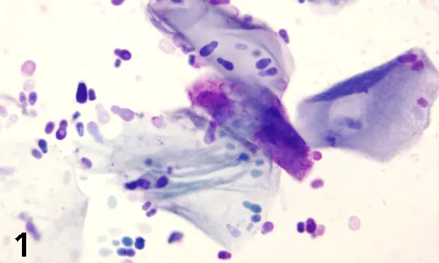
Figure 1. Smear from left ear canal. (modified Wright’s stain; original magnification, 100×)
FoundationPathophysiology. Otitis externa is a common disorder affecting the external ear canal of dogs. Primary causes include parasites (e.g., Otodectes species), allergic disorders (e.g., atopy, food hypersensitivity), foreign bodies, immune-mediated disorders, seborrhea, or endocrinopathies (e.g., hypothyroidism). Predisposing factors include ear anatomy (e.g., pendulous pinna, stenosis of the canal), environmental humidity and heat, swimming or bathing, excessive ear cleaning, or systemic disorders with immune compromise. Factors that perpetuate otitis include bacteria, yeast, other fungal infections, proliferative changes, and treatment errors or reactions.1,2
Clinical signs. Pain, pruritus, reddening or swelling of the pinna and/or external canal, head shaking, odiferous discharge.
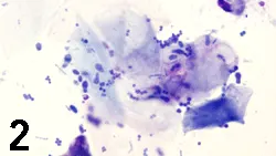
Smear from right ear canal. (modified Wright’s stain; original magnification, 100×)
Diagnosis: Otitis externa, mixed infection
Cytologic Evaluation. The smear from the left ear canal (Figure 1) contained large numbers of squamous epithelial cells and markedly increased numbers of broad-based budding yeasts. The findings were consistent with an overgrowth of Malassezia pachydermatis. The smear from the right ear canal (Figure 2) contained squamous epithelial cells, scattered neutrophils, large numbers of bacteria (cocci, fewer rods), and fewer Malassezia organisms. The findings were consistent with neutrophilic inflammation with a mixed infection.
Diagnosis. Successful treatment of otitis externa requires identification of the underlying cause. Infections cannot reliably be differentiated on the basis of odor or gross appearance of exudates.1 Cytologic examination is critical for accurate diagnosis and for monitoring response to therapy. Ear swabs are easily collected and provide immediate diagnostic information. Swabs should be obtained from both ears because different processes may be present.3 Fine-needle biopsy is required if a mass is present.
Discharge should be collected using a cotton swab before the ear canals are cleaned. Immediately after collection, part of the discharge is placed onto a glass slide and mixed with mineral oil to evaluate for mites. The remainder of the discharge is gently rolled onto another slide that is air-dried and stained with any of the routine cytologic stains (e.g., Diff-Quik, Wright's stain).2,3 Features assessed microscopically include the number and type of bacteria, presence of yeast or other fungal organisms, number and type of inflammatory cells, the presence of excessive cerumen (a combination of the squamous epithelial cells that line the ear canal, keratin, and oily secretions from underlying sebaceous and ceruminous glands), and neoplastic cells.
Cellular Analysis. Squamous epithelial cells are large, individual, angular cells with an absent or pyknotic nucleus and abundant cytoplasm. They are found in ear swabs from healthy dogs, but dogs with otitis may have increased numbers. Because of the high lipid content, the oily secretions in cerumen do not stain with routine cytologic stains and may dissolve during staining.3 Inflammatory cells are rarely found in healthy ear canals.3 Neutrophils and occasionally macrophages occur with infections or neoplasia.
Bacteria & Fungi. Healthy dogs may have occasional bacteria per oil immersion field (100× objective)<sup.1,2sup> Bacteria are identified as rods and/or cocci. Numerous bacteria associated with cerumen in the absence of inflammatory cells suggest colonization of the canal but not necessarily clinical infection. These bacteria are probably an incidental finding, but may contribute to odor and inflammation by causing lipolysis of the cerumen. Bacteria in combination with neutrophils and phagocytized organisms indicate active clinical infection<sup.4sup>
Malassezia pachydermatis is a yeast commonly identified in the ears of healthy dogs as well as those with otitis. Identification of more than nine organisms per high-power field (40× objective) or two to three organisms per epithelial clump indicates that the yeast are contributing to the otitis, even if inflammatory cells are absent.3 Other fungi are rarely associated with otitis in dogs (e.g., Candida, Aspergillus, Microsporum species).
Ask Yourself...
• What would you expect to find on cytologic examination of an ear swab from a healthy dog?• What microscopic features should be evaluated in an ear swab from a dog with otitis?• What microscopic findings are present in the smears from this dog?• What are the distinguishing characteristics of the organisms?
Did You answer...
• Ear swabs from healthy dogs often contain cerumen, a waxy material containing squamous epithelial cells and oily secretions from sebaceous and ceruminous glands. There may be occasional bacteria and small numbers of yeasts (M. pachydermatis). Inflammatory cells are rare.• Before drying, a smear should be evaluated for mites. Another smear should be stained and evaluated microscopically for bacteria, yeasts, other fungal organisms, inflammatory cells, excessive cerumen, and neoplastic cells.• The swab from the left ear contains overgrowth of M. pachydermatis. The swab from the right ear contains a mixed infection (bacteria and yeasts) and neutrophilic inflammation.• M. pachydermatis is a broad-based, budding, nonmycelial yeast that varies from peanut to ellipsoid in shape. Bacteria are smaller and appear as basophilic rods and cocci with routine stains.
Related Articles:Myringotomy & Ear Disease ManagementEvaluating & Managing Otitis Externa/Media
OTITIS EXTERNA • Jennifer S. Thomas
References
1. Management of otitis externa. Rosychuk RAW. Vet Clin North Am Small Anim Pract 24:921-952, 1994.2. The external ear canal. Tyler RD, Cowell RL, Baldwin CJ, et al. In Cowell RL, Tyler RD, Meinkoth JH (eds): Diagnostic Cytology and Hematology of the Dog and Cat, 2nd ed-St. Louis: Mosby, 1999, pp 83-87.3. Cytologic evaluation of otic exudates. Chickering WR. Vet Clin North Am Small Anim Pract 18:773-782, 1988.4. Otitis externa. Greene GE. In Green GE (ed): Infectious Diseases of the Dog and Cat, 2nd ed-Philadelphia: WB Saunders Co, 1998, pp 549-554.