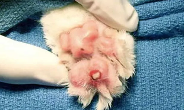
Onychectomy & Tendonectomy
Susanna Hinkle Schwartz, DVM, DACVS, MedVet Medical and Cancer Center for Pets, Cincinnati, Ohio

Profile - Definition
Onychectomy is a very controversial and emotional issue.
This article will not debate the ethics of onychectomy, but will provide information to decrease short- and long-term postoperative complications.
Signalment
Approximately 14.4 million1 cats undergo onychectomy each year; approximately 45% of owned cats are declawed.2
Onychectomy usually involves the front paws of indoor felines (6 months to 3 years of age).
Approximately 50% of cats are declawed at the time of ovariohysterectomy/castration.2
Indications
Scratching furniture and people
Other indications include paronychia (onychomycosis, follicular infection) or nail bed neoplasms (squamous cell carcinoma, melanoma, soft tissue sarcoma, osteosarcoma, mast cell tumors)3,4
Medications
Preoperative Pain Management
Premedication 20 minutes prior to induction has been demonstrated to minimize stress, decrease dose of other anesthetic medications, and lessen postoperative pain.5
Common premedication protocols include:
Hydromorphone and diazepam (0.05–0.2 mg/kg each) or
Buprenorphine (0.01 mg/kg) and acepromazine (0.1 mg/kg) or
Buprenorphine (0.01 mg/kg) and diazepam (0.05–0.2 mg/kg).5
Cats are usually induced with:
Thiopental sodium (8–13 mg/kg) or
Propofol (2–4 mg/kg) or
Ketamine (5.5 mg/kg)/diazepam(0.275 mg/kg).5
Anesthesia is usually maintained with isoflurane or sevoflurane in oxygen.
Preoperative meloxicam (0.3 mg/kg SC) given to cats 15 minutes after premedication and before anesthesia has been shown to result in improved analgesia for 24 hours without clinically relevant adverse effects.6 I use only a preoperative dose of meloxicam at 0.1 mg/kg SC or PO due to the new warning labels on Meloxicam (boehringer-ingelheim.com; see Extra-Label Use of Meloxicam on page 98 of the November issue for more information).
A bupivacaine 4-point ring nerve block of the radial, ulnar, and median nerves can aid in perioperative analgesia (1 mg/kg of a 0.75% solution7 or 0.1–0.2 mL/site of 0.5% bupivacaine, with a total dose not to exceed 5 mg/kg4).
A study performed by Curcio, et al, revealed no difference in discomfort or complication scores between control limbs and limbs receiving nerve blocks.7 In my opinion, a 4-point ring nerve block is minimally invasive and may contribute to multimodal pain management, with cats exhibiting less postoperative pain.
Sites for nerve blocks to the feline forepaw
(A) Extend the carpus and palpate the superficial digital flexor tendon along the palmar aspect of the paw. Block the median nerve with 0.15 mL bupivacaine just medial to the superficial digital flexor tendon. Similarly, block the palmar branches of the ulnar nerve along the lateral superficial digital flexor tendon.(B) Block dorsal digital nerves II to V by inserting the needle from lateral to medial just distal to the carpus. Inject 0.2 mL of bupivacaine as the needle is withdrawn. Block dorsal digital nerve I at the articulation between metacarpal I and II with 0.1 mL of bupivacaine.
Reprinted from Fossum’s Small Animal Surgery, 3rd ed. Fossum TW, Hedlund CS, Hulse DA—Philadelphia: Mosby, 2007, Figure 18-44, with permission.
Postoperative Pain Management
While the patient is hospitalized, IV, IM, or SC injections of butorphanol, hydromorphone, oxymorphone, or buprenorphine are often administered.
In addition, the following medications, administered through routes other than injection, also provide postoperative analgesia:
Buprenorphine: 0.01 to 0.02 mg/kg PO sublingual Q 6 to 8 H8
Butorphanol: 0.5 to 1 mg/kg PO Q 6 to 8 H8
Fentanyl patches: 25 mcg patch applied transdermally8,9
Many studies show that cats are lame after surgery; analgesic medications should be dispensed to owners when the cat is discharged from the hospital.
Laser Onychectomy
A surgical laser may be used to cut the skin and soft tissues of the distal interphalangeal joint. Differences in discomfort and complications between groups of cats on which onychectomy was performed with the blade technique as opposed to carbon dioxide laser were not clinically relevant and were observed only 1 day after surgery.3
In this study, cats in the laser onychectomy group had improved limb function immediately after surgery compared to those in the blade onychectomy group,7 but the improved peak vertical force for the laser group was only observed on days 1 and 2 postoperatively, and was equal between groups by day 3. Holmberg and Brisson found that patients experienced discomfort at 10 days after onychectomy by either laser or blade, but laser onychectomy was associated with less lameness during the first 7 days after surgery.10
Advantages
Tourniquet unnecessary, as the laser vaporizes the tissue and seals the blood vessels and lymphatics11
Avoids tourniquet placement complications (neuropraxia of the radial nerve, tissue ischemia, muscle damage)
Bandages not absolutely necessary, although some surgeons place for 24 hours after surgery
Tissue necrosis subsequent to improper bandage placement does not occur1
Disadvantages
Procedure takes longer to complete11
Safety issues (eg, inhalation of irritating and noxious smoke, laser-induced combustion, and eye and skin burns)12
High cost of purchasing and maintaining laser and smoke evacuation systems
No studies evaluating long-term complications
Surgery
The 2 main surgical options to prevent scratching are tendonectomy and onychectomy.
Tendonectomy
Tendonectomy severs the deep digital flexor tendon to prevent the cat from flexing and extending the third phalanx. After tendonectomy, the claw remains retracted, but the nail continues to grow. Tendonectomy may be less painful than onychectomy,13 but the owner must trim the cat’s nails. There are conflicting results of studies regarding owner satisfaction with tendonectomy versus onychectomy, and there may also be more complications after tendonectomy than onychectomy.14,15
Scrub the feet with a 2% chlorhexidine gluconate solution and saline (0.9% NaCl) or isopropyl alcohol; place the cat in lateral or dorsal recumbency.
Place a tourniquet distal to the elbow to minimize radial, median, and ulnar nerve damage.
Make a small incision on the palmar surface between the second and third phalanx.
Dissect under the shiny white tendon with mosquito hemostats or small scissors; transect and remove approximately 5 mm of the tendon.
Close the skin edges with tissue adhesive or sutures.
Onychectomy
Onychectomy involves removing the third phalanx either using a blade, guillotine-type nail clipper, or surgical laser (see Laser Onychectomy) to cut the supporting soft tissues.
The key to humane onychectomy is pre- and postoperative pain management, nerve blocks, tourniquet application below the elbow, careful soft tissue incisions, and being sure not to cut the pads.
Blade Onychectomy
Aseptic preparation is required for blade onychectomy.
Place a penrose drain tourniquet distal to the elbow joint.
Apply a towel clamp or hemostat to the claw; pull it forward to expose the cuticle-like skin around the nail bed.
Make a circumferential incision at the distal interphalangeal joint using an 11 or 15 blade (the most common blades for this procedure).
After the skin is incised, transect the common digital extensor tendon and dorsal ligaments.
Pass the blade around the contour of the third phalanx to transect the collateral ligaments, followed by the deep digital flexor tendon.
Incise the remaining soft tissues and joint capsule.
Take care to avoid cutting the digital paw pads as this is thought to result in increased pain.
Remove the entire third phalanx to prevent nail or bone regrowth.
Apply a few drops of surgical skin glue between the skin and squeeze closed, or place cruciate sutures (3–0, monofilament absorbable) taking care to avoid the digital pads.
Most surgeons bandage the paws after blade onychectomy.
Guillotine-Type Onychectomy
A guillotine-type nail clipper (Resco nail shears, teclausa.com/resco) can also be used to perform onychectomy. Aseptic preparation is required for this procedure.
Trim the nails to ease nail clipper placement and pass the digit through the nail clipper.
Grasp the claw with a towel clamp or hemostat and extend the claw.
Place the blade at the dorsal joint surface, then lift the claw to deviate the flexor process ventrally. The curved portion of the nail clipper is typically seated in the dorsal joint space between P2 and P3. Apply the blade of the nail clipper to the ventral surface, using the blade to push the distal edge of the digital pad proximally.
Be sure the digital pad is not in the blade and close the blade.
Inspect the third phalanx to ensure that the entire palmar process is removed. Typically, a small fragment of the flexor process of P3, the point at which the digital flexors attach, remains. The key issue is to ensure that the ungual crest (location of germinal tissue) has been completely removed to prevent nail regrowth.
Remove any remaining bone with a scalpel blade or scissors.
Close and bandage as in the blade declaw technique.4
Short-term complications are more common with the blade technique, but long-term complications are more common with guillotine-type nail clipper technique.4
A surgical laser may also be used to cut the skin and soft tissues of the distal interphalangeal joint.
Follow-Up
Patient Monitoring
Remove bandages 12 to 24 hours after declaw. If bleeding persists, replace bandages for an additional 1 to 3 days.
Instruct owners to use shredded paper rather than cat litter and restrict exercise for 1 to 2 weeks after surgery.
Recheck 2 weeks postoperatively to assess the declaw site and remove sutures if present.
Complications
The most common short-term postoperative complications are hemorrhage, pain, and soft tissue swelling.
The most common long-term postoperative complications are claw regrowth, protrusion
of the second phalanx, incisional dehiscence/infection/draining tract, and persistent lameness.
Uncommon long-term complications include radial nerve paralysis from tourniquet use; tissue necrosis from improper bandage placement; and rarely, cystitis,and asthma.1
In General
In my opinion, onychectomy is an appropriate surgery when performed correctly. The key is to complete the procedure in the most humane way possible, including preoperative pain medication; nerve blocks; tourniquet application below the elbow; careful soft tissue incisions, being sure not to cut the pads; and appropriate postoperative pain management. Laser is an appropriate method of performing onychectomy but has not been proven superior to the blade or guillotine-type nail clipper technique.
TX at a Glance
First, attempt to prevent scratching through behavioral training with positive reinforcement. Encourage owners to use scratching posts and nonsurgical treatments, such as Soft Paws (softpaws.com) vinyl caps (placed every 6 to 8 weeks; the cat's nails will still need to be trimmed on a regular basis).
Surgical options include onychectomy and tendonectomy.