Obtaining High-Quality Dental Radiographs
Candice Hoerner, CVT, VTS (Dentistry), Ponderosa Veterinary Hospital, Kalispell, Montana

Part 1 of a 2-part series
Dental disease can cause pain and infection in veterinary patients, but clinical signs may not always be apparent. Dental radiography discloses pathologic changes below the gingival margin and is an important tool for diagnosing and treating hidden disease.
Understanding the Machine
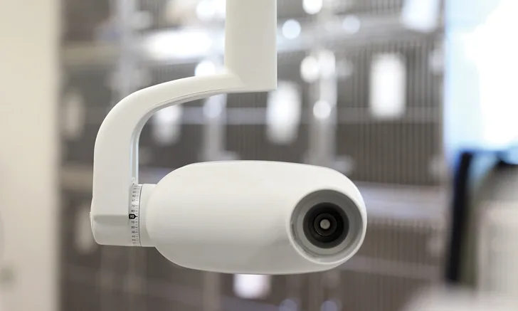
A dedicated dental radiography machine is essential for producing high-quality diagnostic images. Veterinary nurses should be familiar with the different components of the machine to ensure images are captured correctly and efficiently.
Wall-Mounted & Mobile Stand Units
The dental radiography unit can be mounted to the wall or on a mobile stand. A wall-mounted unit is dedicated to a particular dental treatment area, but the varying lengths of the flexible arm can extend to the patient work area and collapse against the wall to save space. The mobile unit is on a wheeled stand that can be moved to different treatment areas and stored where convenient in the practice.1
Tube Head
The tube head is attached to the flexible arm mount and contains the radiography tube, which converts electrical energy from the generator to radiation and heat. The amount of radiation generated is determined by settings for kilovoltage (kV), milliamperage (mA), and exposure (mA/second [mAs]). The radiation produced in the tube is emitted toward the intended target as a controlled beam through the radiography cone or position-indicating device (PID). The PID creates the appropriate focal distance to the plate or sensor. The scale of angulation typically is set on the tube head.2
Control Panel
This allows the user to set the mA, the kilovoltage peak (kVp), and the mAs to produce a properly exposed image. In many units, the kVp and mA are already set, and preset exposure times (in mAs) can be selected depending on the patient species and teeth to be imaged.
Exposure Button
The button is used to obtain the image according to the values specified on the control panel. This button may be located on the control panel or be attached to a stretchable cord.
Digital Plate or Sensor
Most units today are computerized and use plates or sensors instead of standard film to capture digital images. Computed radiography uses photostimulable phosphor plates to capture images, which are scanned and converted into digital radiographs. Plates are available in various sizes and can be erased and reused. Direct radiography uses a sensor connected directly to a computer and provides an image immediately. In veterinary medicine, the size-2 sensor is most commonly available. The flat side of the sensor or the light side of the plate should face the radiography beam.2
Imaging Software
User-friendly image-capture software provides tools for enhancing digital images and templates that streamline the capture process. The templates are organized by tooth type, and the software will automatically proceed to the next preset image once the first image has been exposed. Images can be saved in a database and organized by patient.
Steps for Obtaining High-Quality Dental Radiographs
To minimize patient discomfort and movement and prevent potential bite reflexeswhich can damage the plate or sensoradminister general anesthesia to patients undergoing dental radiography. Sensors and plates should be protected with a sensor sheath cover.
Position the patient according to the teeth being imaged. Using the simplified radiography technique, the patient will be placed in sternal recumbency for the maxillary teeth and dorsal recumbency for the mandibular teeth. This patient positioning technique allows the veterinarian to obtain multiple images in a timely manner with minimal patient movement and repositioning. The patient also can be placed in lateral recumbency, but the simplified angles would not apply and the patient would have to be moved several times.
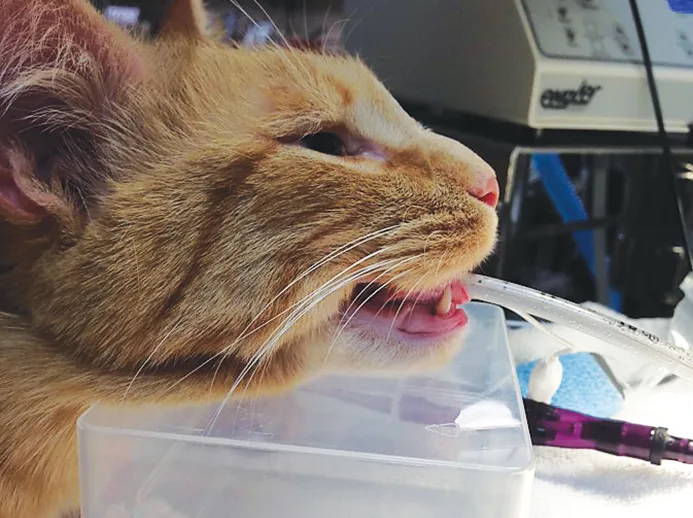
Imaging maxillary teeth: Place the patient in sternal recumbency with the head slightly elevated so the palate is parallel with the table. (See Figure 1.)
Imaging mandibular teeth: Place the patient in dorsal recumbency. If necessary, place a neck bolster or towel under the neck so the ventral surface of the mandible is parallel to the table. (See Figure 2.)

FIGURE 1 To obtain radiographs of maxillary teeth, position the patient in sternal recumbency, and elevate the head so the palate is parallel to the table.Photos courtesy of Candice Hoerner, CVT, VTS (Dentistry)
Determine the best technique based on the patient and teeth being imaged.
Parallel technique requires inserting the plate or sensor into the patients mouth parallel to the tooth being imaged. Position the PID perpendicular to the plate or sensor. The parallel technique is used to obtain images of the caudal mandible.
The bisecting angle technique uses the plane of the tooths long axis and the plane of the plate or sensor to create an angle that is bisected with an imaginary line. The radiography beam is aimed perpendicular to this bisected line. This method produces the most accurate radiographs but can create confusion and can be time consuming until mastered.2
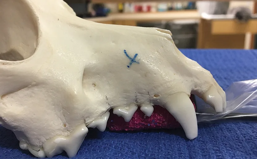
Simplified bisecting angle technique requires placing the plate or sensor in the mouth so the teeth crowns are near the edge of the plate or sensor, parallel to the table, and positioning the PID at a preset vertical angle. (See Figures 3-8.) Only a limited number of angles are needed to obtain most dental radiographs. (Use the parallel technique to obtain images of the mandibular third and fourth premolars and molars.3)

FIGURE 3 When obtaining views of the maxillary canine root apex (X), place the sensor as shown in this model.
Place the plate or sensor in the mouth according to the selected technique.
Set the PID angle according to the selected technique (see Table 1), and press the exposure button to obtain the radiograph.
Recommended Angles & Positions
, This tube head angle slightly elongates the image and thins the zygomatic bone to allow image interpretation. (See Figure 8.)
Dental Radiography Tips
Create a standard sequence (eg, incisors, canines, premolars, molars) for obtaining images.
Use the software templates to move quickly from one image to the next as you move to different teeth throughout the full-mouth sequence.
For large teeth (eg, canines, mandibular first molars), obtain 2 images to capture the entire tooth.
Use gauze or small pieces of foam to help keep the plate or sensor in position.
Wrap a small piece of flexible bandage material over the sensor sheath cover to provide traction and prevent the sensor from slipping out of position.
Position the PID as close as possible to the patient to minimize image distortion.
Make sure the radiography beam is centered on the area or tooth to be radiographed.
Never use sharp objects near the plate or sensor.
Practice taking full-mouth radiographs on every patient to help quickly improve technique.
Conclusion
Radiography is a valuable tool for identifying hidden dental and oral disease and determining appropriate treatment. Nearly 50% of a tooths structure lies below the gingival margin. Teeth with crowns that appear normal can have extensive pathology that only can be detected with radiography. Diagnostic radiographs can be obtained using several techniques, including the simplified bisecting angle technique, which is simple to learn and, with practice, consistently produces high-quality radiographs. Use of dental radiography increases the quality of patient care and enhances the level of service provided by a veterinary practice.