The Not-So-Routine Pyometra Case

An approximately 9-year-old intact female boxer presented with polydipsia, polyuria, anorexia, and mental depression. Abnormal physical examination findings included 7% to 8% dehydration, grossly suppurative vaginal discharge, prominent hyperpigmented nipples, vulvar edema, and hyperpigmentation along the perineum and caudal abdomen (Figures 1 and 2). Laboratory analysis revealed evidence of mild anemia, leukopenia with a mild left shift, and thrombocytopenia. Vaginal cytology was not done.
Abdominal radiographs revealed uteromegaly and an apparent mass lesion just caudal to the right kidney. Thoracic radiographs revealed no abnormalities. On the basis of the hemogram, a bone marrow aspirate was not deemed necessary.
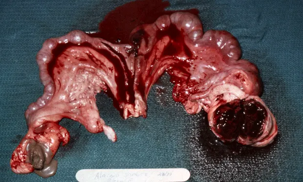
According to the clinical findings, the tentative diagnosis included pyometra and an ovarian mass. Surgery and histopathology confirmed that the right-sided mass was an ovarian granulosa cell tumor and a pyometra (incised after excision to show uterine contents) (Figure 3). The dog also had a left-sided ovarian teratoma (Figure 4). A complete bilateral ovariohysterectomy was done. The dog was treated with intravenous fluids and antibiotics after surgery and made an uncomplicated recovery. A follow-up hemogram performed 2 weeks later revealed normal red and white blood cell counts and a normal platelet count.
Granulosa cell tumors are the most common sex cord tumors, accounting for 50% of these tumors in the dog. Metastasis has been noted in up to 30% of cases according to 1 report. The cells arise from ovarian granulosa and thecal cells, making them capable of secreting estrogen or progesterone. Hyperestrogenism, in turn, can cause myelosuppression, which can lead to aplastic anemia or bone marrow hypoplasia. In addition, prolonged progesterone exposure can predispose to pyometra. Surgery can be curative if metastasis has not occurred.
Teratomas are derived from the primordial germ cells and account for 6% to 12% of canine ovarian tumors. These germ cells can undergo differentiation into 2 or more germ cell layers, with any combination of ectodermal, mesodermal, or endodermal tissues being seen. A metastasis rate as high as 32% has been reported for these tumors. Surgery can be curative if done before metastasis occurs.