A New Technique for Emergency Vascular Access
Steven L. Marks, BVSc, MS, MRCVS, Diplomate ACVIM, North Carolina State University
Rita Hanel, DVM, Diplomate ACVIM & ACVECC, North Carolina State University
Intraosseous catheterization involves placing a needle or catheter into the medullary cavity of a bone.
Intraosseous catheterization can be used as a route for emergency vascular access in patients with marked hypotension, in pediatric patients, or during cardiopulmonary and cerebral resuscitation. This technique has been previously described in the veterinary literature as an option when IV access is not available,1-3 and complements information provided in an earlier Clinician’s Brief article.4
EZ-IO Technique
The EZ-IO (vidacare.com) system provides a new technique for performing intraosseous or bone marrow catheterization. The technique is ideally used for the short term until alternate vascular access is available.
EZ-IO is marketed as an emergency intraosseous infusion system for critically ill humans in the prehospital setting (eg, battlefield, ambulance) or for in-hospital emergency resuscitation. It is reported to have excellent efficacy and serves as a bridge from initial stabilization to central line placement in critically ill people.
System Components
The EZ-IO infusion system includes:
A hand-held, battery-powered drill, which allows rapid placement (< 10 seconds) of a purpose-made intraosseous catheter
Catheters that are constructed of stainless steel with a plastic hub and contain a styleted needle with a cutting tip for rapid intraosseous access. A black line marked 5 mm from the needle hub assists with placement.
Three sizes of catheters are available for use with this system
Pediatric size: 15-gauge (1.8-mm diameter), 15-mm long
Adult size: 15-gauge, 25-mm long
A 45-mm-long needle that has not been used in veterinary patients
Once the stylet is removed, the remaining plastic hub, which contains a Luer lock connector, can be attached to a t-port, syringe, or any standard fluid administration set.-Although these catheters are marketed for single use, they can be sterilized via an autoclave; we have reused them up to 4 times without adverse procedural effects.
Catheterization Sites
Recommended anatomic sites for intraosseous catheterization include the trochanteric fossa of the femur, crest or wing of the ileum, tibial tuberosity, or greater tubercle of the humerus. We suggest using the greater tubercle of the humerus or the tibial tuberosity for the EZ-IO system.
Step-by-Step: EZ-IO Intraosseous Catherization
What You Will Need
Clipper with clean blades
Antiseptic cleansing solution
2% lidocaine solution
3-mL syringe
22-gauge hypodermic needle
# 11 scalpel blade
EZ-IO infusion system
Step 1
Place the patient in lateral recumbency and identify the greater tubercle of the humerus. The landmarks for localization include the scapular spine and the acromion.


Step 2
Shave and aseptically prepare the anatomic site. Infiltrate the skin and subcutis with approximately 0.25 to 0.5 mL of 2% lidocaine and make a small stab incision. In some emergency cases, such as cardiopulmonary cerebral resuscitation, local anesthetic infiltration and the stab incision are skipped.
Author Insight
Once the tip of the needle is placed in the periosteum of the greater tubercle, apply forward pressure to keep the needle from slipping off the cortical bone.
Step 3
Load the intraosseous catheter onto the power driver. Push the tip of the needle through the skin and place it into the periosteum of the greater tubercle. Apply forward pressure to make sure the needle does not slip off the cortical bone. Depress the power button of the drill and drill the catheter into the bone.


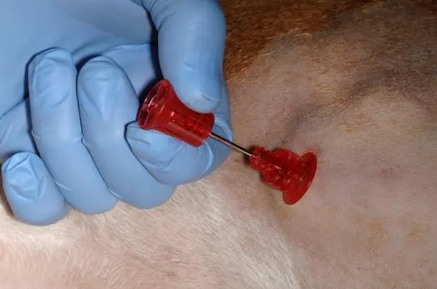
Step 4
Once the catheter is placed, firmly implant the hub. You should be able to move the leg with the hub of the catheter. Remove the stylet. Placement can be confirmed by aspiration of bone marrow through the catheter. Aspiration may result in some minor discomfort, causing the patient to react.


Step 5
A t-port, syringe, or fluid administration set can be attached for administration of emergency drugs or crystalloid or colloid solutions.

Step 6
Solutions can be administered at rates similar to those used intravenously, but the rates may need to be slower in smaller patients. Drug dosages for intraosseous administration are the same as intravenous dosages. Pain may be associated with high rates of fluid infusion, which may be reduced by the infusion of lidocaine through the catheter (1–2 mg/kg, 2% solution).
Author Insight
Drug dosages for intraosseous administration are the same as intravenous dosages.