Nasal Discharge in a Labrador Retriever
Peter S. Chapman, BVetMed (Hons), DECVIM-CA, DACVIM, MRCVS, Veterinary Specialty & Emergency Center, Levittown, Pennsylvania

History
A 12-year-old, neutered male Labrador retriever was presented for evaluation of a 4-month history of chronic nasal discharge. The discharge was unilateral from the left side and had progressed from clear to mucopurulent with a blood-tinged component. The dog had been sneezing more frequently over the same time period but was otherwise healthy. Body weight was stable, and no significant findings had been noted in the previous medical history.
Physical Examination
A small amount of epistaxis from the left nostril was evident at the time of examination, but no facial pain or asymmetry was apparent. Although airflow was subjectively normal through both nostrils when assessed with a piece of cotton wool, heavy stertorous breathing was noted at rest, which worsened with excitement. The remainder of the examination was unremarkable.
Differential Diagnoses
Nasal neoplasia: Based on patient signalment, unilateral nature of the discharge, and presence of epistaxis, neoplasia was considered the most likely diagnosis at the time of the initial examination.
Fungal rhinitis: This condition also can be unilateral. The character of nasal discharge can vary from a typical profuse mucopurulent discharge to overt epistaxis.
Foreign body: Nasal foreign bodies may typically cause unilateral nasal discharge but are usually associated with an acute onset and severe bouts of paroxysmal sneezing.
Chronic rhinitis: This common condition usually involves bilaterally symmetric signs with mucopurulent discharge; it is less common to see a sanguineous component to the discharge, but persistent inflammation and sneezing may occasionally lead to low-grade epistaxis.
Dental disease: Dental disease associated with the tooth roots can occasionally cause unilateral signs of nasal disease.
Systemic disease: Coagulopathies and hypertension can cause epistaxis, but a mucopurulent discharge would not be expected.
Diagnostic Plan
Initial screening diagnostics, including serum chemistry profile, hematology panel, prothrombin time, activated partial thromboplastin time, and thoracic radiography were all unremarkable.
Complete evaluation of the nasal cavity in patients with chronic nasal discharge includes diagnostic imaging, endoscopic examination, and collection of samples for histopathologic or cytologic analysis.
Imaging
Skull radiography (open-mouth ventrodorsal, skyline frontal sinus, lateral skull views): Radiography can show soft tissue masses and/or turbinate destruction, but rarely can the underlying cause be definitively identified.
Advanced imaging: Because computed tomography (CT) and magnetic resonance imaging (MRI) show considerably more detail of the nasal cavity than skull radiography does, these imaging modalities are preferred. CT affords greater detail of bony lesions, while MRI better details soft tissue lesions. Both nasal neoplasia and fungal rhinitis are associated with significant changes to the bony turbinates, which are more evident on CT.1 As a result, the higher cost and potentially longer procedure time associated with MRI are generally not justified in patients with nasal disease.
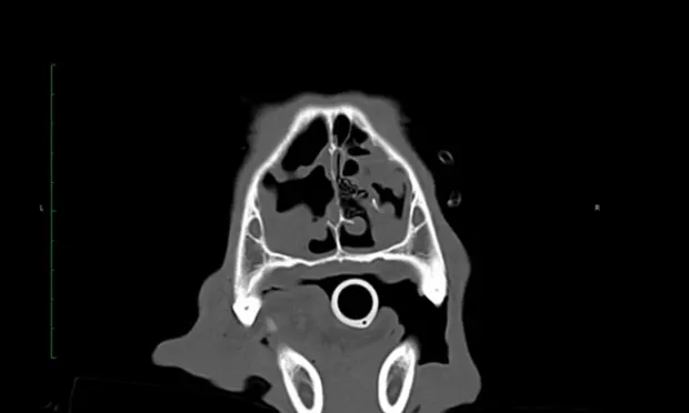
In this patient, CT (Video 1) revealed changes consistent with fungal rhinitis, including destruction of nasal turbinates and lesser flat bone destruction without evidence of a soft tissue mass (Figure 1) and irregular noncontrast-enhanced material in the frontal sinus, along with hyperostosis of the frontal bone (Figure 2).

Figure 1.
Two CT slices at different levels through the nasal cavity showing cavitated lesions secondary to turbinate destruction (D) in the left nasal cavity. There are milder changes in the right nasal cavity. (L = left, R = right)
For comparison, Video 2 shows CT of a dog with chronic rhinitis. Some patchy soft tissue/fluid density can be seen in the region of the nasal turbinates. However, no significant bony destruction is evident, nor is there any opacity within the frontal sinuses or frontal bone sclerosis.
Rhinoscopy
Rhinoscopy was performed to visualize lesions and collect samples to confirm a diagnosis of fungal rhinitis. A complete rhinoscopic examination should begin by placing a flexible endoscope retroflexed over the soft palate to view the choanae and then inserting a rigid or flexible endoscope to evaluate the rostral nasal cavity. Flexible antegrade rhinoscopy is technically feasible only in the largest canine patients.
Rhinoscopy allows visualization of only about 50% of the nasal cavity and none of the paranasal sinuses.2 Thus it is advantageous to use both rhinoscopy and advanced imaging to fully evaluate the nasal cavity. In large patients, an otoscope can be placed to visualize the most rostral portions of the nasal cavity, but the diagnostic yield of this technique is very low, with only the most rostral portions of the nasal cavity being acccessible.2
In this patient, the choanae appeared normal on a retroflex view (Figure 3). On rigid antegrade rhinoscopy, marked destructive changes were seen in the left nasal cavity, along with localized accumulation of debris caudally, consistent with a fungal plaque (Figures 4 and 5). No mass or foreign body was seen. The right side was unremarkable.
Diagnosis: Fungal Rhinitis
The most common causal agent of canine fungal rhinitis is Aspergillus fumigatus, a ubiquitous soil saprophyte that is introduced to the nasal cavity by inhalation. This disease is most common in young to middle-aged mesocephalic or dolichocephalic breeds. Signs can vary from chronic mucopurulent discharge to more severe findings, including life-threatening epistaxis and seizures attributed to destruction of the cribriform plate. Ulceration or depigmentation of the nasal planum is a common finding on physical examination but was not noted in this patient. The prognosis for fungal rhinitis is fair to good, although successful treatment is challenging and relapses may occur.
Treatment
This patient was treated via endoscopic debridement, followed by temporary trephination of both frontal sinuses. The sinuses were debrided and lavaged before being infused with 1% clotrimazole solution and packed with a depot of 1% clotrimazole cream.5 Fungal rhinitis can also be treated noninvasively by occluding the nostrils and nasopharynx with Foley catheters, instilling clotrimazole solution, and rotating the patient through ventral, dorsal, left, and right recumbent positioning during a 1-hour contact time.6 However, this procedure may result in treatment failure because of inadequate debridement of or poor drug penetration into the frontal sinus.4 Oral drugs are generally ineffective because the fungus resides on the mucosal surface and does not invade underlying tissues.4
Dr. Chapman is affiliated with Veterinary Specialty & Emergency Center (VSEC), with offices located in Philadelphia and Levittown, Pennsylvania. He specializes in his board certification area of internal medicine, with focus on endocrinology, gastroenterology, and hematology.