Mitral Valve Disease in a Dog
Teresa DeFrancesco, DVM, DACVIM (Cardiology), DACVECC, North Carolina State University College of Veterinary Medicine

OSCAR, A 14-YEAR-OLD, CASTRATED DACHSHUND MIX, presented with a 12 dayhistory of progressive cough, intermittent tachypnea with increased respiratory effort, and lethargy, and no interest in eating or drinking in the previous 12 hours. His 3-year history of heart murmur was characterized on imaging as myxomatous mitral valve (MV) disease with moderate left atrial enlargement; enalapril therapy was started at diagnosis. On examination, he was anxious and shaking. His heart and respiratory rates were 170 beats per minute and 60 breaths per minute, respectively. He was normothermic and normotensive.


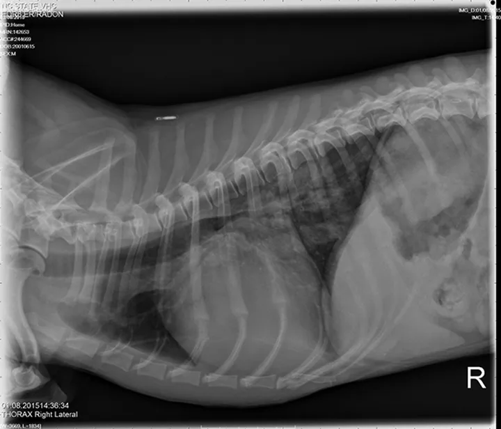
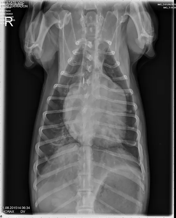
A grade IV/VI systolic heart murmur was detected with a point of maximal intensity over the left apical region; heart rhythm was regular, and pulses were strong and synchronous. Lung sounds were increased, with no crackles or wheezes. Thoracic radiographs showing progressive left atrial and ventricular enlargements, mildly enlarged pulmonary veins, a moderate patchy unstructured interstitial pattern in the right caudal lung lobe, and a mild unstructured interstitial pattern in the left caudal lung lobe were consistent with pulmonary edema. Caudal mainstem bronchi were compressed on lateral projections secondary to the cardiomegaly and left atrial enlargement. Radiographic findings were compatible with left-sided congestive heart failure (CHF) secondary to MV disease.
Lateral and DV views showing mild CHF in a dachshund with mitral valve disease.
ACE = angiotensin-converting enzyme, CHF = congestive heart failure, MV = mitral valve, RAAS = renin-angiotensin-aldosterone system