Lissamine Green Staining for Tear Film Deficiency Evaluation
Anja Welihozkiy, DVM, DACVO, BluePearl Veterinary Partners, Sarasota, Florida
In the Literature
Smith SM, Holt E, Aguirre GD. Conjunctival staining with lissamine green as a predictor of tear film deficiency in dogs. Vet Ophthalmol. 2020;23(4):624-631.
The Research …
Canine tear film deficiencies are commonly diagnosed in veterinary medicine and include both quantitative (keratoconjunctivitis sicca [KCS]) and qualitative deficiencies.1 Qualitative tear film deficiencies result from abnormal mucin or lipid distribution/production of the precorneal tear film and cause tear film instability with increased tear evaporation and poor tear coverage of the ocular surface. Both quantitative and qualitative deficiencies can cause ocular irritation, as evidenced by conjunctival hyperemia; discharge; blepharospasm due to discomfort; palpebral, conjunctival, and corneal inflammation; and, in chronic cases, corneal pigmentation.
A Schirmer tear test (STT) is a quantitative measurement of aqueous tear production: >15 mm/minute is considered normal in dogs, and <15 mm/minute, along with clinical signs, is considered diagnostic for KCS. Tear film breakup time (TFBUT) and rose bengal staining have been used clinically to diagnose qualitative tear film deficiency. TFBUT involves fluorescein staining and is used to evaluate the corneal ability to maintain a homogenous tear covering through measurement of the time between dye application and breakup into smaller pools. Normal TFBUT in dogs is >20 seconds; a shorter TFBUT (often <5 seconds) is considered diagnostic for qualitative tear film deficiencies.2 Rose bengal dye stains devitalized cells and healthy cells not covered by an appropriate mucin film; therefore, this dye can be used as an indicator for poor precorneal tear film coverage.3 Recently, lissamine green dye has become more popular in both human and veterinary medicine due to favorable properties (ie, less ocular irritation, less cytotoxicity, better commercial availability) as compared with rose bengal dye.4
This study evaluated whether an easy-to-use grading scale using lissamine green staining (LGS) to evaluate for qualitative tear film deficiency could be used in dogs. Staining strips were wetted with 1 to 2 drops of sterile eyewash and touched once to the ventral bulbar conjunctiva of each eye in 54 dogs. The eyelids were closed and opened to ensure complete coverage. This was followed by immediate evaluation of the bulbar conjunctiva using 10× magnification and the cobalt blue filter of a slit-lamp biomicroscope.
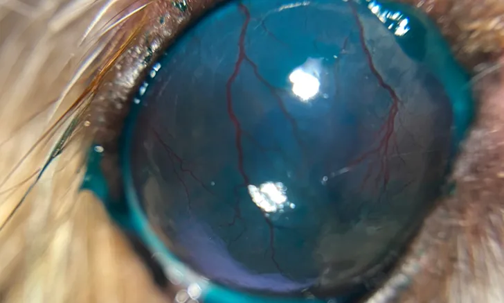
A previously described ocular staining score used in human ophthalmology was modified for this use,5 and 3 subjective grades of temporal conjunctival LGS were assessed based on stain intensity. Grade 0 indicated little to no stain retention on the temporal bulbar conjunctiva (Figure 1), grade 1 indicated mild stain retention, grade 2 indicated moderate stain retention (Figure 2), and grade 3 indicated diffuse stain retention.
There was a significant association between an increase in LGS grade and a decrease in STT readings (<15 mm/minute) and between a more rapid TFBUT and a higher LGS grade. In addition, a higher LGS grade was significantly associated with brachycephalic skull conformation.

LGS grade 0. Little to no stain retention can be seen.


LGS grade 2. Moderate stain retention is present.
… The Takeaways
Key pearls to put into practice:
Increased LGS correlates with increased severity of tear film deficiency in dogs.
During canine ocular examination, a normal STT measurement with an evident LGS indicates qualitative tear film deficiencies.
LGS for evaluation of canine tear film deficiency is easy to perform, with little additional time required.
You are reading 2-Minute Takeaways, a research summary resource presented by Clinician’s Brief. Clinician’s Brief does not conduct primary research.