Lethargy & GI Distress in a Puppy

History and Signalment
Bandit, a castrated Yorkshire terrier (8 months of age), was presented on emergency with a 2-day history of lethargy, vomiting, and diarrhea. He was recently adopted from a rescue organization and had a history of a portosystemic shunt with surgical cellophane banding performed 4 months earlier. He received a liver-protective diet with puppy food; he received no medications.
Physical Examination
At examination, Bandit appeared depressed but was alert and responsive. He showed pale and icteric mucous membranes, was tachycardic (HR, 180 bpm), and was found to have a previously unreported grade III/VI heart murmur. His femoral pulses were hyperdynamic, and his heart rhythm was regular. His abdomen was painful on palpation. His rectal temperature was normal (101°F), and he was eupneic with normal lung sounds.
Diagnostics
Pending CBC, serum biochemistry profile, and coagulation profile results, PCV and TP diagnostics were performed. Bandit was found to be profoundly anemic with a PCV of 14% and TP of 8 mg/dl. The serum in the hematocrit tube was hemolyzed, and a slide agglutination test was negative.
The CBC showed a total WBC count of 41,320; a mature leukocytosis was present. The HCT was 12%, there were 17 nRBCs/100 WBCs, the anemia was highly regenerative with a reticulocyte count of 22% (total reticulocytes of 437,000/µL; range, 6400–81,500/µL), mild spherocytosis, and 15% Heinz Bodies. The serum biochemistry profile showed a total bilirubin of 6.5 mg/dl (normal, <0.3 mg/dl). The coagulation profile was abnormal with a PT of 8.1 seconds (normal, 6.2–7.7 seconds) and a PTT of 57.2 seconds (normal, 9.8–14.6 seconds).

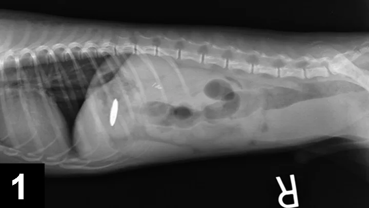
Abdominal radiographs (Figure 1) showed an apparently metal object in the stomach.
Figure 1. Radiograph showing foreign body.
Diagnosis
Based on signs, blood test abnormalities, and radiograph findings, zinc toxicity from ingestion of a penny was suspected.
Treatment
Bandit received a 4-hour transfusion of packed RBCs and fresh frozen plasma simultaneously, using the same donor for each unit. After two hours, he was more cardiovascularly stable, and he was anesthetized for endoscopic retrieval of the apparently metal foreign body. The foreign body was retrieved from the stomach.
Outcome
Bandit was monitored in ICU on IV fluid therapy at 4 ml/kg/hr (ie, Plasmalyte A) and gastric protectants (ie, pantoprazole at 1 mg/kg IV q24h, sucralfate at 250 mg PO q8h) for 48 hours. Intermittent heart rate, respiratory rate, body temperature, mentation, and PCV monitoring was also performed in the ICU. His anemia stabilized to 22%, he was much more mentally alert, and his total bilirubin continued to decrease during hospitalization to a discharge value of 2.6 mg/dl. He was discharged on day 3. He was rechecked 5 days afterward and was found to have a normal serum biochemistry profile (including resolution of his hyperbilirubinemia), a PCV of 27%, and normal examination findings. He was reportedly eating well at home with normal activity.
Zinc Toxicity
Zinc toxicity has been reported in dogs, usually as a result of ingestion of U.S. pennies minted after 1982 (before 1982, pennies consisted primarily of copper). Other zinc-containing foreign bodies include some nuts and bolts, zippers, and zinc oxide creams. When zinc-containing pennies are ingested, the gastric acid liberates the zinc and allows for intestinal absorption to occur. The zinc can also cause significant corrosive GI irritation, so therapy with gastric protectants is highly recommended. Oxidative damage occurs to the RBCs, causing the formation of Heinz bodies and severe, intravascular hemolysis. Coagulation abnormalities are also often noted, likely from the severity of RBC damage and, potentially, a direct effect of the zinc on coagulation proteins.
To verify the diagnosis of zinc toxicity in this dog, zinc levels in the blood could have been performed; however, zinc toxicity was highly suspected based on radiographic identification of a radio-opaque foreign body and was confirmed on penny removal. In cases of zinc toxicity, serial blood levels can be performed to follow resolution of the disease.1
Chelation therapy has been reported in dogs, but there is controversy with its use (ie, calcium EDTA), as the chelated apparently metal may actually be absorbed more easily from the GI tract and also may induce nephrotoxicity. Removal of the zinc source causes toxic levels to decline rapidly, and with supportive care, many dogs recover.2
Because zinc toxicity can mimic immune-mediated hemolytic anemia, all patients presenting with signs consistent with immune-mediated hemolytic anemia (IMHA) should have abdominal radiography performed to ensure zinc ingestion is not the underlying cause.3 Removal of the zinc foreign body, treatment with blood and plasma transfusions as needed, and administration of gastric protectants is indicated. This toxicity can be fatal; however, prognosis can be good with immediate removal of the zinc foreign body, transfusions as needed, and ICU care.