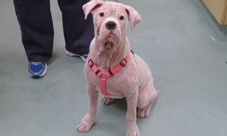
Juvenile Generalized Demodicosis in a Dog
Andrew Rosenberg, DVM, DACVD, Animal Dermatology & Allergy Specialists, White Plains, New York, and Riverdale, New Jersey

A 6-month-old spayed American pit bull terrier crossbreed is presented for evaluation of severe pruritus and multifocal areas of alopecia and erythema. When the owners rescued the dog 2 months prior, the dog was pruritic around the eyes and licked its paws. The skin has since become progressively worse and the dog has gotten progressively more pruritic. The owners have been feeding a strict limited-ingredient diet without improvement.
Physical examination reveals crusts on the lateral cervical region and diffuse erythroderma. All distal limbs and both dorsal and ventral aspects of all paws are severely erythematous and crusted. The ventral abdomen and inguinal region have multifocal papules and pustules. An impression smear reveals intracellular and extracellular cocci bacteria (too numerous to count) with streaming neutrophils. Deep skin scrapings from the right forepaw and dorsal muzzle reveal 15 live adult Demodex canis mites, 2 juvenile Demodex canis mites, and 5 Demodex canis eggs per slide.
Conclusion
Regardless of the treatment choice for this patient’s generalized demodicosis, miticidal treatment should continue until multiple consecutive skin scrapes obtained a month apart are completely negative for all evidence of Demodex spp mites. Antibiotic therapy should continue until at least 1 week beyond clinical resolution of all superficial bacterial skin infection. Because there is a genetic component causing a predisposition to the development of juvenile generalized demodicosis, affected dogs should not be bred.