Iris Freckles, Nevi, & Melanosis
Gil Ben-Shlomo, DVM, PhD, DACVO, DECVO, Iowa State University
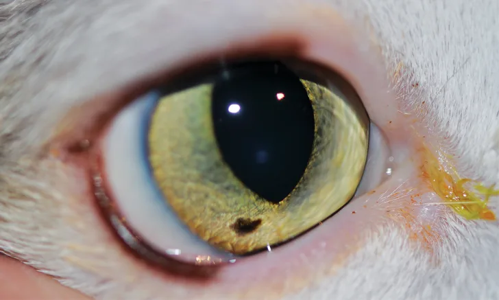
Figure 1 Iris freckle in a cat. A discrete, focal area of flat hyperpigmentation is present in the ventral portion of the iris.
The terms iris freckle, nevus, and/or focal melanosis have been used interchangeably in many instances in the veterinary literature, although they have not been characterized in veterinary patients. Until there are further studies in veterinary patients, these conditions can be differentiated based on the human literature. Iris freckles (ie, focal melanosis; Figure 1) are flat and affect only the iris surface, whereas iris nevi cause focal thickening of the iris stroma and can be slightly raised from the iris surface (Figure 2).
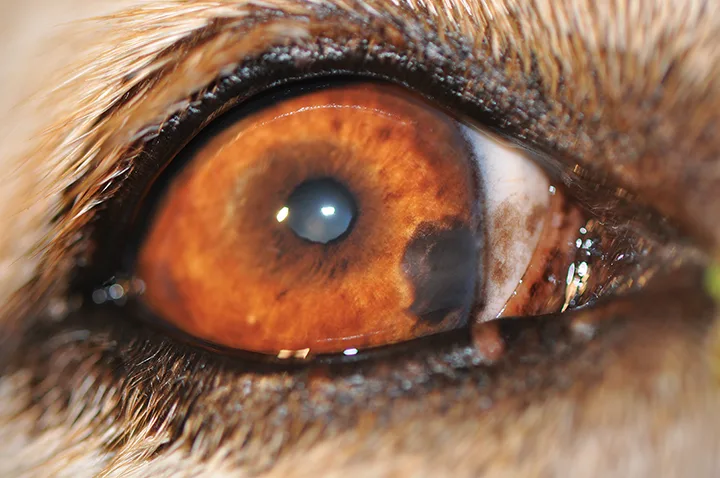
Iris nevus in a dog. A well defined hyperpigmented lesion that is slightly raised from the surface of the iris can be noted in the medial aspect of the iris.
Clinical Signs of Iris Freckles & Nevi
Iris freckles and nevi are focal areas of hyperpigmentation on the iridal surface. Although iris freckles and nevi have been studied extensively in humans, there is little information in the veterinary literature on these conditions in dogs and cats.1,2 In humans, iris freckles are the most common melanocytic iridal lesion and appear to be discrete, superficial colonies of atypical—but benign—melanocytes that are seen primarily in elderly patients.3 It has been suggested that iris freckles in humans may be induced by sunlight exposure,4 and iris nevi are thought to be congenital, caused by a melanocytic acceleration during embryogenesis, and present at a young age.3 Iris freckles and nevi are not associated with other ocular clinical signs (eg, inflammation, increase of intraocular pressure).
Clinical Signs of Iris Melanosis
Melanosis is a condition characterized by dark pigmentary deposits.5 In iris melanosis, the melanocytes are limited to the anterior surface of the iris. Iris melanosis may be focal or multifocal, and multifocal areas may coalesce to form diffuse iris melanosis. In cats, diffuse iris melanosis is usually a unilateral, progressive condition (Figure 3).6 Chronic uveitis may also cause iris melanosis7 and should be addressed if present. In Cairn terriers, golden retrievers, and boxers, ocular melanosis can also cause pigmentation and thickening of the iris (Figure 4). Ocular melanosis is an inherited condition characterized by ocular pigment proliferation, as well as pigment dispersion and deposit in other ocular structures (eg, sclera, episclera, posterior segment), with poor long-term prognosis for the eye and vision.8
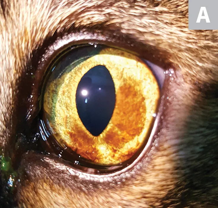
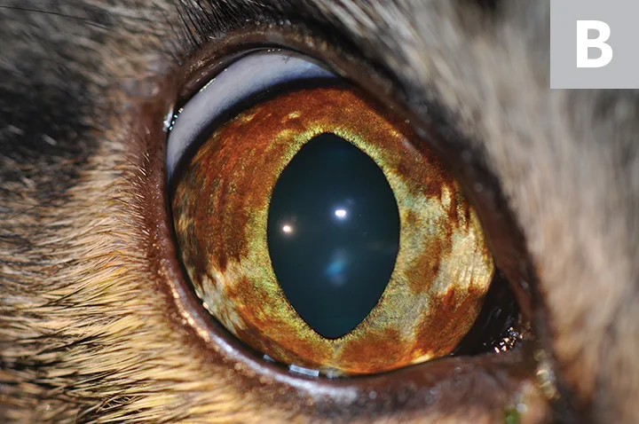
A relatively light diffuse iris melanosis in a cat approaching the iridocorneal angle at the 7 o’clock position (A). A more progressed iris melanosis approaching the iridocorneal angle at almost 360° (B). The pigmentation is denser and occupies a larger area of the iris as compared with the patient in Figure A.
Diagnosis
Diagnosis of iris freckles, nevi, and diffuse iris melanosis is based on clinical signs, as described previously. However, in cats, differentiation between diffuse iris melanosis and a unique form of diffuse iris melanoma cannot be determined based on clinical appearance alone, and only histologic evaluation can confirm diagnosis.6,9 This can complicate the decision to remove a visual, nonpainful eye, as diffuse melanosis is benign and does not pose a risk to the patient; however, early enucleation in cats with diffuse iris melanoma can prevent premature death, as metastasis occurs in 20% to 25% of these patients (see Anterior Uveal Melanocytic Neoplasia).9
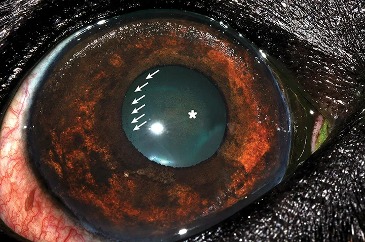
Diffuse iris melanosis in the iris of a dog with ocular melanosis. Pigment dispersion on the anterior lens capsule can be noted both peripherally (arrows) and centrally (asterisk). Examination with a slit lamp revealed numerous pigmented cells floating in the anterior chamber.
Treatment
Iris freckles, nevi, and melanosis do not require treatment. The presence—or lack—of ophthalmologic changes (eg, iris thickening, dyscoria, invasion of the pigment to the iridocorneal drainage angle, changes to the iris architecture, pigment dispersion, involvement of the ciliary body, increased intraocular pressure) may be associated with malignant pigmentary tumors and can aid the decision to enucleate an eye. Thus, patients with iris hyperpigmentation should be closely monitored, ideally by a veterinary ophthalmologist, with advanced diagnostic techniques such as slit lamp biomicroscopy, gonioscopy, and tonometry. The potential malignancy of iridal pigmentary lesions should be discussed with the owner. The decision to enucleate an eye with diffuse pigmentary changes can be difficult, as lesions may be benign and focal areas of hyperpigmentation may exist for months to years before irregular iris masses develop.9
Laser treatment for suspected diffuse iris melanoma in cats has recently been suggested to effectively delay tumor progression10 but remains controversial due to a lack of controlled studies and the difficulty in differentiating benign diffuse iris melanosis from melanoma.
In dogs and other species with iris melanosis due to chronic uveitis,7 uveitis should be treated, and a complete diagnostic investigation should be performed to identify and treat the underlying cause.