Intraosseous Catheterization: A Life-Saving Tool
Elisa M. Mazzaferro, MS, DVM, PhD, DACVECC, Cornell University Veterinary Specialists

Intraosseous catheterization involves placing a needle into a bone.1-4 This procedure should be considered when vascular access is not possible or cannot be performed in adequate time due to cardiac arrest, hypovolemic shock, patient anatomy, presence of skin wounds or thrombosis over proposed sites of intravenous catheterization, or very small patient size. The technique is technically simple to perform, requires no specialized equipment or tools, and can make the difference between life and death in small animals.
Contraindications
Relative contraindications include fracture of the proposed catheter site, bacterial infection or sepsis, skin wound or infection over the proposed site of catheterization, or pneumatic bone in avian species. In avian species, however, placement of a catheter into pneumatic bone can be used to facilitate administration of supplemental oxygen.4
Rate of Infusion
Rate of fluid infusion is directly proportional to diameter of the needle, placement of the needle, whether bevel of the catheter is placed against the bony cortex, and whether needle is partially occluded with bony debris. Fluid can flow with gravity alone, at a maximum rate of 11 mL/min; however, this rate is much slower in extremely small patients. When the catheter is placed under 300 mmHg of pressure, flow can increase to 24 mL/min. In some cases, rapid infusion can increase patient discomfort.
Indications for Intraosseous Catheter Placement
Extremely small body size
Patient anatomy (eg, very short limbs)
Exotic species
Vascular collapse
Hypotension
Severe dehydration
Hypovolemic shock
Hypothermia
Inaccessible IV catheter site
Wounds, edema, or infection over proposed sites
Thrombosis of vessels
Obesity
Step-by-Step: Intraosseous Catheterization

Step 1A.
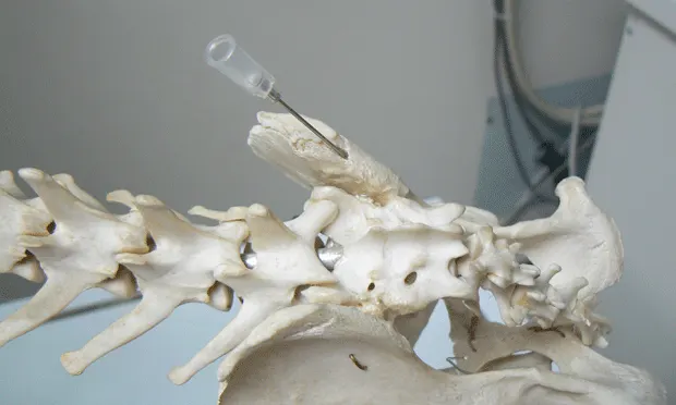
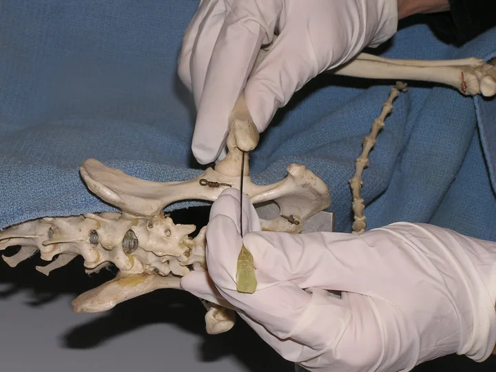
Locations for intraosseous catheter placement include the trochanteric fossa of the femur (A and B), the wing of the ileum (C), the proximal humerus (D), and the tibial tuberosity (E). The trochanteric fossa is easy to catheterize; the tibial tuberosity is an excellent place for catheterization of exotic species, including reptiles.
Figure 1A. The trochanteric fossa of the femur.
Contraindications for Intraosseous Catheter Placement
Skin wound and/or infection over proposed catheter site
Fracture of bone intended for catheterization
Pneumatic bones
Sepsis
Metabolic bone disease
Potential Complications of Intraosseous Catheter Placement
Osteomyelitis
Damage to epiphysis
Fluid leakage into subcutaneous tissues
Edema
Editor’s note: This article was originally published in May 2009 as “Intraosseous Catheterization: An Often Underused, Life-Saving Tool.”