TABLE OF CONTENTS
The following image gallery depicts eyes of patients with decreased intraocular pressure (IOP) and increased IOP due to primary and secondary mechanisms.
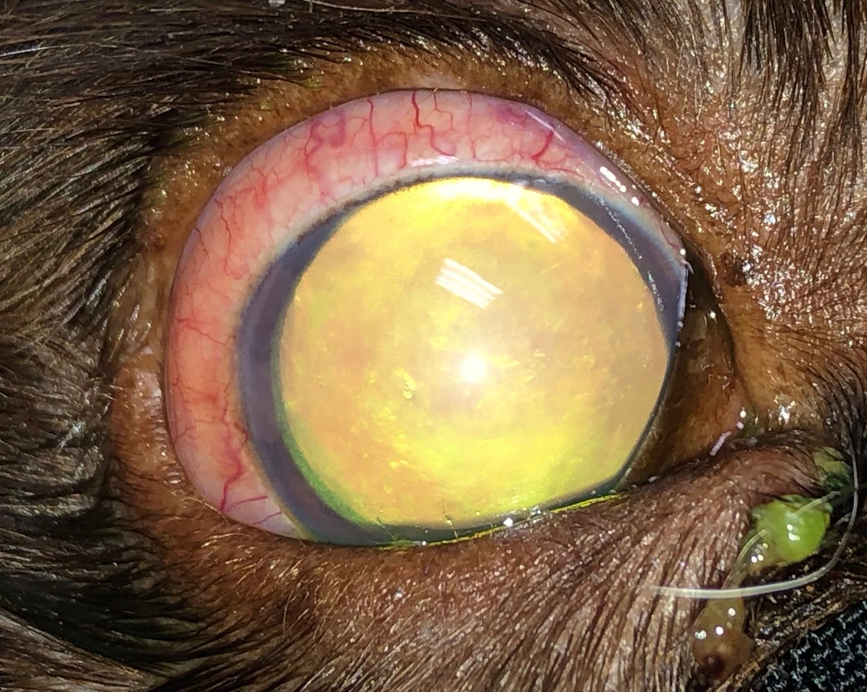
Figure 1
1 / 5
Figure 1
Right eye of an 8-year-old neutered male American cocker spaniel with acute primary glaucoma. The episcleral injection and mydriatic pupil can be observed. IOP was 48 mm Hg (normal, 10-20 mm HG).
Listen to the Podcast
Sponsor message; content continues afterward
Trusted content.
Tailored to you.
For free.
Create an account for free.
Want free access to the #1 publication for diagnostic and treatment information? Create a free account to read full articles and access web-exclusive content on cliniciansbrief.com.