Inappropriate Urination in a Neutered Cat
Carla Barstow, DVM, MS, DACT, Highland Pet Hospital, Lakeland, Florida

History
Bandit, a 3-year-old neutered male cat, was presented for inappropriate urination. He had a history of nonobstructive feline idiopathic cystitis, which had been treated successfully, according to the pet owner, with a course of antibiotics. The last episode was 6 months prior. The owner reported that Bandit sprayed on vertical surfaces (eg, side of the couch) approximately once a week and demonstrated highly energetic and sometimes aggressive behavior (eg, “He just seemed wild”). Bandit had been adopted at one year of age from a local shelter, where he was reported to have been neutered before adoption.
Physical Examination
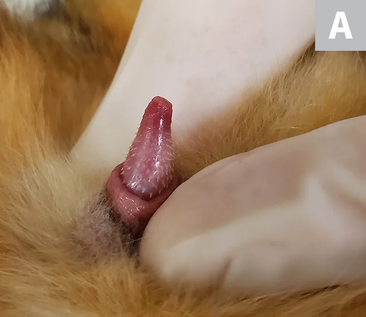
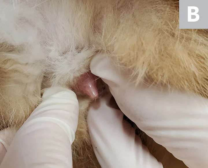
On examination, Bandit was bright, alert, and responsive. His BCS was 3/5. Vital parameters and thoracic auscultation were within normal limits. The bladder was small and nonpainful on abdominal palpation. No testicles were noted in the scrotum. The penis was extruded and examined, and penile spines were noted (Figure 1). The remainder of the physical examination was within normal limits.


Penile spines on the patient (A). For comparison, a penis without spines from a neutered male cat (B)
Diagnosis
Causes of feline inappropriate urination can be medical (eg, feline lower urinary tract disease, feline idiopathic cystitis, urinary calculi), hormonal (eg, intact status, exogenous hormonal therapy, adrenal tumor), or related to litter box aversion or urine marking.1
Because penile spines, which are testosterone-dependent, were noted on physical examination, this case of inappropriate urination was most likely due to hormonal causes. Differential diagnoses for penile spines in a presumably neutered male cat are retained testicle(s), exogenous testosterone exposure, or a sex hormone-producing adrenal tumor.
Access to topical hormonal creams in the household was ruled out, and, because Bandit was an indoor-only cat, exogenous testosterone exposure was unlikely. Adrenal tumors are rarely reported in cats.2 Retained testicular tissue can be diagnosed via imaging, hormone testing, or exploratory laparotomy (see Diagnosing Retained Testicular Tissue).
The owner wanted to ensure testicular tissue was present before surgery was performed, so an anti-Müllerian hormone (AMH) test was submitted to the laboratory, and results confirmed the presence of testicular tissue.
DIAGNOSING RETAINED TESTICULAR TISSUE
Imaging
Ultrasonography can help identify the presence and location of retained testicle(s) and rule out an adrenal tumor.
Hormone Testing
Baseline testosterone levels can be diagnostic indicators; however, testosterone may be low even in the presence of testicular tissue because of seasonality6 or because testosterone is secreted in pulsatile bursts.
Gonadotropin-releasing hormone (GnRH) or human chorionic gonadotropin (hCG) stimulation tests can offer an accurate and more complete evaluation of testosterone levels. During these tests, baseline blood testosterone is drawn, GnRH or hCG is administered, and a second blood sample is drawn to evaluate testosterone concentration 2 to 4 hours later. A marked increase in testosterone levels indicates presence of testicular tissue.
An AMH level test can help evaluate for presence of testicular tissue and is preferred because the assay has 100% specificity and sensitivity in detecting neuter status in cats.7 Testing for AMH only requires a single blood draw (vs multiple draws for stimulation tests).
Surgery
Abdominal exploratory surgery can be performed to find the retained testicle(s).
Diagnosis: Retained Testicular Tissue
Treatment & Long-Term Management
Spraying behavior can be controlled via removal of the hormone source. It is highly recommended to neuter patients that exhibit this behavior to help minimize or even eliminate marking behavior.
Nonsurgical options can be considered. Melatonin implants have been used to nonsurgically temporarily control fertility; although these have been shown to decrease sperm production, they do not affect testosterone levels.3 Because marking behaviors in cats can be associated with elevated testosterone levels due to the patient being intact, this treatment option most likely would not be an effective solution in this patient. Deslorelin, a synthetic GnRH-agonist, can also induce temporary infertility in treated patients. Urine marking disappeared in study cats within 10 weeks of treatment, and the effects lasted ≈20 months.4 Of note, deslorelin is only FDA-approved for use in ferrets in the United States.
Bandit was scheduled for exploratory laparotomy based on the positive AMH results. His inguinal regions were carefully palpated while he was under general anesthesia; inguinal testicular tissue was not palpated. Because the presence of one or both testicles was unconfirmed, anesthetized ultrasonography was used to identify testicular tissue presence andlocation. Both testicles were identified in the caudal abdomen.
Retained abdominal testicles can be located anywhere from the caudal pole of the kidney to the scrotum. It is important to locate the ductus deferens and trace it until the testicle is located.
A caudal midline abdominal incision was made; the right testicle was easily located next to the bladder. The ductus deferens was identified on the left side and traced caudally. The bladder was then retroflexed to identify the left testicle, which was positioned near the inguinal ring. Both testicles were removed and submitted for histopathology. The abdomen was closed using a routine 3-layer closure.
Recovery from anesthesia was uneventful, and histopathology confirmed complete removal of both retained testicles.
TREATMENT AT A GLANCE
Physical examination in cases of inappropriate urination in male cats should include investigation for penile spines.
Neutering is indicated to decrease urine spraying in male cats that have retained testicular tissue.
Ultrasonography to determine location of retained testicle(s) can help guide surgical planning.
Histopathology should be used to confirm complete removal of testicular tissue.
Prognosis & Outcome
Bandit was presented for examination 6-weeks postoperatively, and penile spines were no longer present. In general, regression of penile spines can take up to 6 weeks after removal of the testosterone source. The owner reported a decrease in urine marking, and Bandit’s demeanor was much calmer than previously noted.
Urine spraying is typical in sexually intact cats, which are often hormonally driven to spray. Neutering reduces the influence of hormones on this behavior; however, 10% of neutered cats may still continue urine marking and spraying.5
TAKE-HOME MESSAGES
Feline spraying of vertical surfaces can be related to hormonal causes.
In cases of inappropriate urination, intact status should be ruled out.
An AMH assay can be used to rapidly determine intact status when spines are present on the penis.
Neutering or removal of the hormonal influence may help decrease further inappropriate urination.