Imaging of Myxomatous Valvular Disease
Myxomatous valvular degeneration (MVD) or endocardiosis is the most common acquired heart disease in dogs. MVD usually affects the mitral valve, and auscultation typically reveals a holosystolic murmur that is heard best at the left apex. Although a rough correlation has been found between murmur intensity and MVD severity in the Cavalier King Charles spaniel,1 severity of mitral regurgitation should not be assessed by murmur intensity alone.
Many factors influence regurgitant murmur intensity, including the pressure difference between the ventricle and the atrium; size, shape, and location of the regurgitant orifice; direction of the regurgitant jet; blood viscosity; size, shape, and contractility of the left ventricle; and the sound transmittng characteristics of the lung and chest wall.
Although a diagnosis of MVD can frequently be based on signalment and murmur, evaluation of the severity of regurgitation should include thoracic radiography, echocardiography, or both.

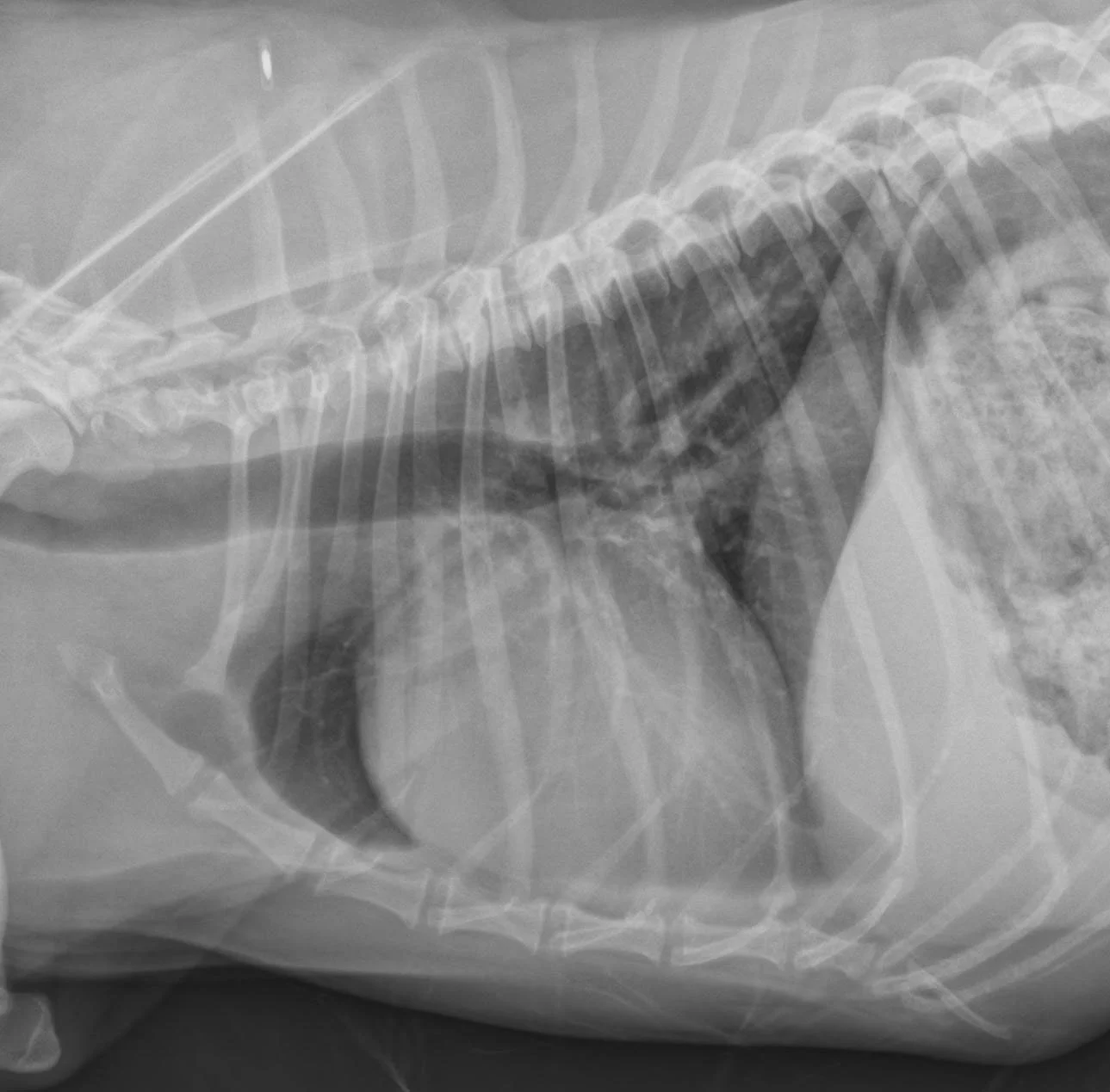
Right lateral (Figures 1 and 2) and dorsoventral (Figures 3 and 4) radiographs of 2 dogs with grade 3/6, holosystolic, left apical murmurs. Dog 1 (Figures 1 and 3) has mild left atrial enlargement; dog 2 (Figures 2 and 4) has a severely enlarged left atrium.