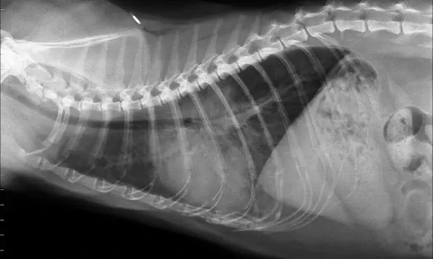
Image Quiz: Feline Emergency Respiratory Distress
Lisa L. Powell, DVM, DACVECC, BluePearl Veterinary Partners, Eden Prairie, Minnesota

Several underlying disease processes can result in respiratory distress, including primary lung disease, heart failure, pleural space disease, traumatic injury, and upper airway disease.
Because stress and handling can exacerbate respiratory disease, cats with dyspnea should initially be placed in an oxygen cage or administered flow-by oxygen on arrival. Respirations should be observed from afar, as the pattern can often indicate which part of the respiratory system is affected. Prolonged inspiratory and/or expiratory effort should be noted, as should any noises audible without the aid of a stethoscope; these are usually associated with upper airway obstruction. Thoracic and tracheal auscultation should be performed, along with an abbreviated physical examination, depending on patient stability.
Because cardiomyopathies are the most common form of heart disease in cats, many cats in respiratory distress associated with congestive heart failure do not have a heart murmur. Hypothermia is often noted in cats in heart failure. In addition, arrhythmias are often caused by primary heart disease.
Common Signs
Cats with upper airway obstruction often display stridorous breathing in addition to respiratory distress. Lower airway disease such as asthma is typically associated with wheezing and expiratory difficulty. Pulmonary disease is often associated with increased bronchovesicular sounds, pulmonary crackles, and respiratory effort during both inspiration and expiration. The breathing pattern associated with pleural space disease is often characterized by increased abdominal effort, inspiratory dyspnea, and passive expiration. Decreased cranial thoracic compressibility can be noted on physical examination in cats with an anterior mediastinal mass.
A quick thoracic ultrasound (thoracic focused assessment with sonography for trauma [TFAST]) can be performed, allowing for rapid diagnosis of pleural effusion, and can reveal evidence of a pneumothorax. If either is present, thoracocentesis should be performed. If the TFAST is negative for significant pleural space disease, thoracic radiographs should be obtained. Care must be taken to not overly stress the cat during radiography, and oxygen should be administered during the procedure.
The following images exhibit commonly diagnosed emergency feline respiratory diseases. Match the image with the description of the disease process.