Image Gallery: Positioning for Abdominal Radiographs
Janet Paquette, AS, LVMT, University of Tennessee

Abdominal radiographs are an important diagnostic tool for determining intra-abdominal disease or abnormalities in the canine or feline patient. Abdominal radiographs are indicated for anything from vomiting/diarrhea to pregnancy checks, but visualization of any structure is dependent on correctly positioned patients. This article demonstrates proper positioning for routine and specialty abdominal radiographs in dogs and cats.
A complete abdominal series should include right and left lateral views as well as a ventrodorsal view. Since motion artifact can be a problem in abdominal radiographs, the use of adequate sedation is recommended to decrease motion. Sedation will also help with patient comfort. Radiographs should be taken during an expiratory pause so the diaphragm is in a cranial position and not compressing the abdominal contents.1
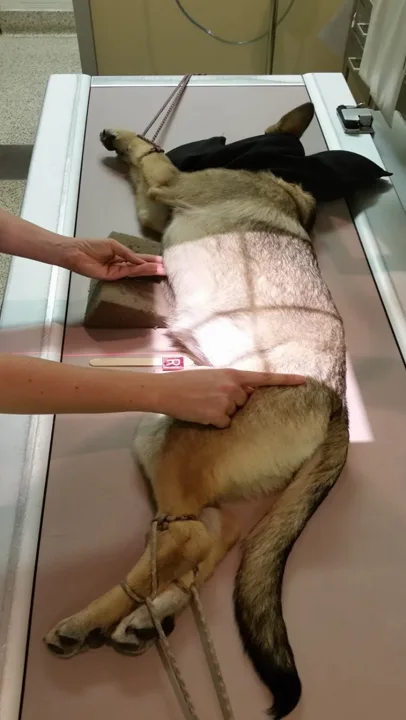
Proper positioning, patient comfort, and minimum radiation exposure are important when imaging the abdomen. Positioning aids (ie, V-trough, sponges, ropes, tape, sedation) will help achieve this, and prevent the need for restraint.

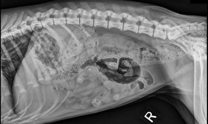
FIGURE 1: Lateral Abdominal View
The lateral abdominal view includes the entire diaphragm cranially and the greater trochanter caudally. Position the cranial edge of the collimator light 2 to 3 fingerbreadths cranial to the xiphoid process. Align the caudal edge of the light at the level of the greater trochanter. Center the beam over the caudal aspect of the 13th rib.1 Using soft ropes or tape, pull the thoracic limbs cranially and pelvic limbs caudally. Pulling the hind limbs caudally will help eliminate superimposition of the femoral muscles over the caudal portion of the abdomen.1 Collimate to include the patient’s dorsal and ventral borders in the radiograph.
This article originally appeared in the July 2016 web issue of Veterinary Team Brief.