The following image gallery depicts eyes of patients with decreased intraocular pressure (IOP) and increased IOP due to primary and secondary mechanisms.
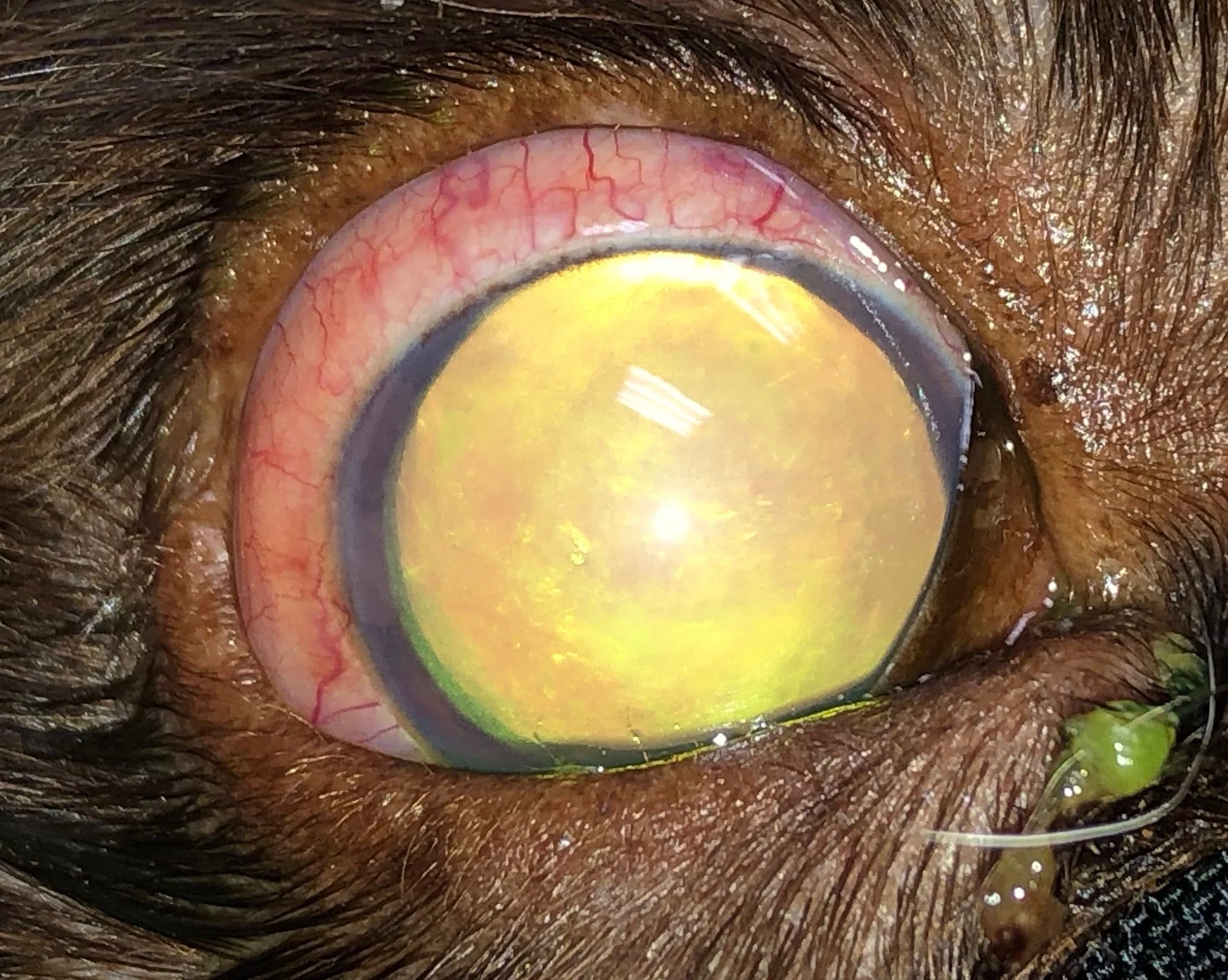
Figure 1
1 / 5
Figure 1
Right eye of an 8-year-old neutered male American cocker spaniel with acute primary glaucoma. The episcleral injection and mydriatic pupil can be observed. IOP was 48 mm Hg (normal, 10-20 mm HG).